 Movie
Movie Controller

[English] 日本語
 Yorodumi
- PDB-3stw: Crystal Structure of tomato Methylketone Synthase I complexed wit... -
+ Open data

- Basic information
| Entry | Database: PDB / ID: 3stw | ||||||
|---|---|---|---|---|---|---|---|
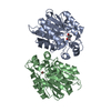
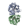
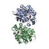
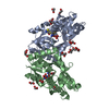
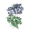
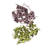
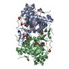
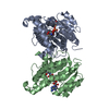
| Title | Crystal Structure of tomato Methylketone Synthase I complexed with 2-tridecanone | ||||||
 Components Components | Methylketone synthase 1 | ||||||
 Keywords Keywords | HYDROLASE / methylketone / alpha/beta hydrolase / decarboxylase | ||||||
| Function / homology |  Function and homology information Function and homology informationmethyl salicylate esterase activity / methyl indole-3-acetate esterase activity / methyl jasmonate esterase activity / salicylic acid metabolic process / jasmonic acid metabolic process Similarity search - Function | ||||||
| Biological species |  | ||||||
| Method |  X-RAY DIFFRACTION / SYNCHROTRON / MOLECULAR REPLACEMENT / Resolution: 2.31 Å X-RAY DIFFRACTION / SYNCHROTRON / MOLECULAR REPLACEMENT / Resolution: 2.31 Å | ||||||
 Authors Authors | Auldridge, M.E. / Austin, M.B. / Noel, J.P. | ||||||
 Citation Citation | Journal: Plant Cell / Year: 2012 Title: Emergent Decarboxylase Activity and Attenuation of alpha/beta-Hydrolase Activity during the Evolution of Methylketone Biosynthesis in Tomato. Authors: Auldridge, M.E. / Guo, Y. / Austin, M.B. / Ramsey, J. / Fridman, E. / Pichersky, E. / Noel, J.P. | ||||||
| History |
|
- Structure visualization
| Structure viewer | Molecule: MolmilJmol/JSmol |
|---|
- Downloads & links
-Download
| PDBx/mmCIF format | 3stw.cif.gz | 115 KB | Display | PDBx/mmCIF format |
|---|---|---|---|---|
| PDB format | pdb3stw.ent.gz | 88 KB | Display | PDB format |
| PDBx/mmJSON format | 3stw.json.gz | Tree view | PDBx/mmJSON format | |
| Others |  Other downloads Other downloads |
-Validation report
| Arichive directory | https://data.pdbj.org/pub/pdb/validation_reports/st/3stwftp://data.pdbj.org/pub/pdb/validation_reports/st/3stw | HTTPS FTP |
|---|
-Related structure data
| Related structure data | 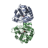 3sttC 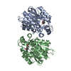 3stuC  3stvC  3stxC  3styC 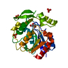 7yasS C: citing same article ( S: Starting model for refinement |
|---|---|
| Similar structure data |


-Links
 PDBj
PDBj

- Assembly
| Deposited unit | 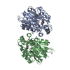
| ||||||||
|---|---|---|---|---|---|---|---|---|---|
| 1 |
| ||||||||
| Unit cell |
|
-Components
| #1: Protein | Mass: 29130.938 Da / Num. of mol.: 2 Source method: isolated from a genetically manipulated source Source: (gene. exp.) Gene: MKS1 / Plasmid: pHis9GW / Production host:  #2: Chemical |   Mass: 196.329 Da / Num. of mol.: 2 / Source method: obtained synthetically / Formula: C13H24O Mass: 196.329 Da / Num. of mol.: 2 / Source method: obtained synthetically / Formula: C13H24O#3: Water | ChemComp-HOH / |  Mass: 18.015 Da / Num. of mol.: 222 / Source method: isolated from a natural source / Formula: H2O Mass: 18.015 Da / Num. of mol.: 222 / Source method: isolated from a natural source / Formula: H2O |
|---|
-Experimental details
-Experiment
| Experiment | Method: X-RAY DIFFRACTION / Number of used crystals: 1 |
|---|
- Sample preparation
| Crystal | Density Matthews: 2.66 Å3/Da / Density % sol: 53.74 % |
|---|---|
| Crystal grow | Temperature: 277 K / Method: vapor diffusion, hanging drop / pH: 6.5 Details: 0.1M PIPES-Na+, 17% (w/v) PEG 8000, 0.3M NaBr, 2mM dithiothreitol, overnight soak in 1mM 2-tridecanone, pH 6.5, vapor diffusion, hanging drop, temperature 277K |
-Data collection
| Diffraction | Mean temperature: 100 K | ||||||||||||||||||||||||||||||||||||||||||||||||||||||||||||||||||||||||||||||||||||||||||||||||
|---|---|---|---|---|---|---|---|---|---|---|---|---|---|---|---|---|---|---|---|---|---|---|---|---|---|---|---|---|---|---|---|---|---|---|---|---|---|---|---|---|---|---|---|---|---|---|---|---|---|---|---|---|---|---|---|---|---|---|---|---|---|---|---|---|---|---|---|---|---|---|---|---|---|---|---|---|---|---|---|---|---|---|---|---|---|---|---|---|---|---|---|---|---|---|---|---|---|
| Diffraction source | Source: SYNCHROTRON / Site: ALS  / Beamline: 8.2.1 / Wavelength: 1 Å / Beamline: 8.2.1 / Wavelength: 1 Å | ||||||||||||||||||||||||||||||||||||||||||||||||||||||||||||||||||||||||||||||||||||||||||||||||
| Detector | Type: ADSC QUANTUM 315r / Detector: CCD / Date: Aug 26, 2007 | ||||||||||||||||||||||||||||||||||||||||||||||||||||||||||||||||||||||||||||||||||||||||||||||||
| Radiation | Monochromator: double crystal Si(111) / Protocol: SINGLE WAVELENGTH / Monochromatic (M) / Laue (L): M / Scattering type: x-ray | ||||||||||||||||||||||||||||||||||||||||||||||||||||||||||||||||||||||||||||||||||||||||||||||||
| Radiation wavelength | Wavelength: 1 Å / Relative weight: 1 | ||||||||||||||||||||||||||||||||||||||||||||||||||||||||||||||||||||||||||||||||||||||||||||||||
| Reflection | Resolution: 2.31→20 Å / Num. all: 30189 / Num. obs: 26600 / % possible obs: 99.6 % / Observed criterion σ(F): 0 / Observed criterion σ(I): -3 / Redundancy: 4.6 % / Biso Wilson estimate: 27.452 Å2 / Rmerge(I) obs: 0.11 / Net I/σ(I): 13.52 | ||||||||||||||||||||||||||||||||||||||||||||||||||||||||||||||||||||||||||||||||||||||||||||||||
| Reflection shell | Diffraction-ID: 1
|
- Processing
| Software |
| ||||||||||||||||||||||||||||||||
|---|---|---|---|---|---|---|---|---|---|---|---|---|---|---|---|---|---|---|---|---|---|---|---|---|---|---|---|---|---|---|---|---|---|
| Refinement | Method to determine structure: MOLECULAR REPLACEMENT Starting model: 7YAS Resolution: 2.31→20 Å / Occupancy max: 1 / Occupancy min: 1 / σ(F): 0 / Stereochemistry target values: MAXIMUM LIKELIHOOD
| ||||||||||||||||||||||||||||||||
| Solvent computation | Bsol: 28.0365 Å2 | ||||||||||||||||||||||||||||||||
| Displacement parameters | Biso max: 74.02 Å2 / Biso mean: 24.82 Å2 / Biso min: 2 Å2
| ||||||||||||||||||||||||||||||||
| Refinement step | Cycle: LAST / Resolution: 2.31→20 Å
| ||||||||||||||||||||||||||||||||
| Refine LS restraints |
| ||||||||||||||||||||||||||||||||
| Xplor file |
|
