 Movie
Movie Controller

[English] 日本語
 Yorodumi
- PDB-6vtz: Structure of a thiolation-reductase di-domain from an archaeal no... -
+ Open data

- Basic information
| Entry | Database: PDB / ID: 6vtz | ||||||
|---|---|---|---|---|---|---|---|
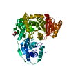
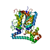
| Title | Structure of a thiolation-reductase di-domain from an archaeal non-ribosomal peptide synthetase | ||||||
 Components Components | Non-ribosomal peptide synthetase | ||||||
 Keywords Keywords | OXIDOREDUCTASE / non-ribosomal peptide synthetases / peptide carrier protein / reductase domain | ||||||
| Function / homology |  Function and homology information Function and homology informationamino acid activation for nonribosomal peptide biosynthetic process / secondary metabolite biosynthetic process / ligase activity / phosphopantetheine binding / cytoplasm Similarity search - Function | ||||||
| Biological species |  Methanobrevibacter ruminantium (archaea) Methanobrevibacter ruminantium (archaea) | ||||||
| Method |  X-RAY DIFFRACTION / SYNCHROTRON / MOLECULAR REPLACEMENT / Resolution: 2.65 Å X-RAY DIFFRACTION / SYNCHROTRON / MOLECULAR REPLACEMENT / Resolution: 2.65 Å | ||||||
 Authors Authors | Deshpande, S. / Lott, J.S. / Lee, T.V. | ||||||
| Funding support |  New Zealand, 1items New Zealand, 1items
| ||||||
 Citation Citation | Journal: J.Biol.Chem. / Year: 2021 Title: Structural characterization of a PCP-R didomain from an archaeal nonribosomal peptide synthetase reveals novel interdomain interactions. Authors: Deshpande, S. / Altermann, E. / Sarojini, V. / Lott, J.S. / Lee, T.V. | ||||||
| History |
|
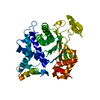
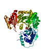
- Structure visualization
| Structure viewer | Molecule: MolmilJmol/JSmol |
|---|
- Downloads & links
-Download
| PDBx/mmCIF format | 6vtz.cif.gz | 91.3 KB | Display | PDBx/mmCIF format |
|---|---|---|---|---|
| PDB format | pdb6vtz.ent.gz | 65.6 KB | Display | PDB format |
| PDBx/mmJSON format | 6vtz.json.gz | Tree view | PDBx/mmJSON format | |
| Others |  Other downloads Other downloads |
-Validation report
| Arichive directory | https://data.pdbj.org/pub/pdb/validation_reports/vt/6vtzftp://data.pdbj.org/pub/pdb/validation_reports/vt/6vtz | HTTPS FTP |
|---|
-Related structure data
| Related structure data | 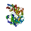 6vtjC  4f6cS C: citing same article ( S: Starting model for refinement |
|---|---|
| Similar structure data |


-Links
 PDBj
PDBj


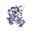
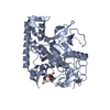
- Assembly
| Deposited unit | 
| ||||||||
|---|---|---|---|---|---|---|---|---|---|
| 1 |
| ||||||||
| Unit cell |
|
-Components
| #1: Protein | Mass: 56423.746 Da / Num. of mol.: 1 Source method: isolated from a genetically manipulated source Source: (gene. exp.) Methanobrevibacter ruminantium (strain ATCC 35063 / DSM 1093 / JCM 13430 / OCM 146 / M1) (archaea)Strain: ATCC 35063 / DSM 1093 / JCM 13430 / OCM 146 / M1 / Gene: mru_0351 / Production host:  |
|---|---|
| #2: Water | ChemComp-HOH /  Mass: 18.015 Da / Num. of mol.: 5 / Source method: isolated from a natural source / Formula: H2O Mass: 18.015 Da / Num. of mol.: 5 / Source method: isolated from a natural source / Formula: H2O |
-Experimental details
-Experiment
| Experiment | Method: X-RAY DIFFRACTION / Number of used crystals: 1 |
|---|
- Sample preparation
| Crystal | Density Matthews: 3.59 Å3/Da / Density % sol: 65.77 % |
|---|---|
| Crystal grow | Temperature: 293.15 K / Method: vapor diffusion, hanging drop / pH: 7.5 Details: Crystals were grown by vapour diffusion, hanging drop method. Drop volume was 2 micro-litre (1+1) equilibrated against 500 micro-litre of reservoir volume containing 0.1 M MOPS/Na-HEPES ...Details: Crystals were grown by vapour diffusion, hanging drop method. Drop volume was 2 micro-litre (1+1) equilibrated against 500 micro-litre of reservoir volume containing 0.1 M MOPS/Na-HEPES buffer at pH 7.5, 15% PEG MME and 14% PEG 20K as precipitants in the presence of 0.02M each of L-Na Glutamate, DL-Alanine, Glycine, DL-Lysine HCl, DL-Serine as additives. The concentration of protein was 30 mg/ml in HEPES buffer at pH 7.5 containing 150 mM NaCl |
-Data collection
| Diffraction | Mean temperature: 100 K / Serial crystal experiment: N |
|---|---|
| Diffraction source | Source: SYNCHROTRON / Site: Australian Synchrotron  / Beamline: MX2 / Wavelength: 0.953 Å / Beamline: MX2 / Wavelength: 0.953 Å |
| Detector | Type: DECTRIS EIGER X 16M / Detector: PIXEL / Date: Jul 19, 2017 |
| Radiation | Protocol: SINGLE WAVELENGTH / Monochromatic (M) / Laue (L): M / Scattering type: x-ray |
| Radiation wavelength | Wavelength: 0.953 Å / Relative weight: 1 |
| Reflection | Resolution: 2.65→46.291 Å / Num. obs: 23800 / % possible obs: 99.9 % / Redundancy: 10.72 % / Biso Wilson estimate: 75.77 Å2 / CC1/2: 0.996 / Rrim(I) all: 0.221 / Net I/σ(I): 7.07 |
| Reflection shell | Resolution: 2.65→2.83 Å / Num. unique obs: 8693 / CC1/2: 0.338 |
- Processing
| Software |
| |||||||||||||||||||||||||||||||||||||||||||||
|---|---|---|---|---|---|---|---|---|---|---|---|---|---|---|---|---|---|---|---|---|---|---|---|---|---|---|---|---|---|---|---|---|---|---|---|---|---|---|---|---|---|---|---|---|---|---|
| Refinement | Method to determine structure: MOLECULAR REPLACEMENT Starting model: 4F6C Resolution: 2.65→46.291 Å / SU ML: 0.4 / Cross valid method: THROUGHOUT / σ(F): 1.34 / Phase error: 26.24 / Stereochemistry target values: ML
| |||||||||||||||||||||||||||||||||||||||||||||
| Solvent computation | Shrinkage radii: 0.9 Å / VDW probe radii: 1.11 Å / Solvent model: FLAT BULK SOLVENT MODEL | |||||||||||||||||||||||||||||||||||||||||||||
| Displacement parameters | Biso max: 131.02 Å2 / Biso mean: 73.5979 Å2 / Biso min: 30 Å2 | |||||||||||||||||||||||||||||||||||||||||||||
| Refinement step | Cycle: final / Resolution: 2.65→46.291 Å
| |||||||||||||||||||||||||||||||||||||||||||||
| LS refinement shell | Refine-ID: X-RAY DIFFRACTION / Rfactor Rfree error: 0 / % reflection obs: 100 %
|
