- PDB-6vsx: X-ray crystal structure of the C-terminal domain of Bacillus subt... -
+
Open data
ID or keywords:
Loading...
-
Basic information
Entry
Database: PDB / ID: 6vsx
Title
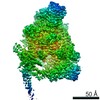
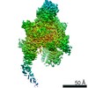
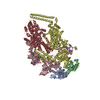
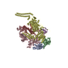
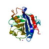
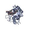
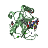
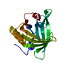
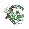
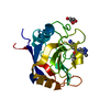
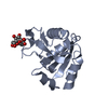
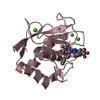
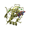
X-ray crystal structure of the C-terminal domain of Bacillus subtilis RNA polymerase binding helicase HelD
Components
DNA helicase
Keywords
TRANSCRIPTION / RNA polymerase / Helicase
Function / homology
Function and homology information
transcription elongation factor activity / recombinational repair / 3'-5' DNA helicase activity / DNA helicase activity / Hydrolases; Acting on acid anhydrides; Acting on acid anhydrides to facilitate cellular and subcellular movement / DNA helicase / hydrolase activity / ATP hydrolysis activity / DNA binding / ATP binding / cytosol Similarity search - Function
National Institutes of Health/National Institute of General Medical Sciences (NIH/NIGMS)
GM131860
United States
Citation
Journal: Nat Commun / Year: 2020 Title: Mycobacterial HelD is a nucleic acids-clearing factor for RNA polymerase. Authors: Tomáš Kouba / Tomáš Koval' / Petra Sudzinová / Jiří Pospíšil / Barbora Brezovská / Jarmila Hnilicová / Hana Šanderová / Martina Janoušková / Michaela Šiková / Petr Halada / ...Authors: Tomáš Kouba / Tomáš Koval' / Petra Sudzinová / Jiří Pospíšil / Barbora Brezovská / Jarmila Hnilicová / Hana Šanderová / Martina Janoušková / Michaela Šiková / Petr Halada / Michal Sýkora / Ivan Barvík / Jiří Nováček / Mária Trundová / Jarmila Dušková / Tereza Skálová / URee Chon / Katsuhiko S Murakami / Jan Dohnálek / Libor Krásný / Abstract: RNA synthesis is central to life, and RNA polymerase (RNAP) depends on accessory factors for recovery from stalled states and adaptation to environmental changes. Here, we investigated the mechanism ...RNA synthesis is central to life, and RNA polymerase (RNAP) depends on accessory factors for recovery from stalled states and adaptation to environmental changes. Here, we investigated the mechanism by which a helicase-like factor HelD recycles RNAP. We report a cryo-EM structure of a complex between the Mycobacterium smegmatis RNAP and HelD. The crescent-shaped HelD simultaneously penetrates deep into two RNAP channels that are responsible for nucleic acids binding and substrate delivery to the active site, thereby locking RNAP in an inactive state. We show that HelD prevents non-specific interactions between RNAP and DNA and dissociates stalled transcription elongation complexes. The liberated RNAP can either stay dormant, sequestered by HelD, or upon HelD release, restart transcription. Our results provide insights into the architecture and regulation of the highly medically-relevant mycobacterial transcription machinery and define HelD as a clearing factor that releases RNAP from nonfunctional complexes with nucleic acids.
In the structure databanks used in Yorodumi, some data are registered as the other names, "COVID-19 virus" and "2019-nCoV". Here are the details of the virus and the list of structure data.
Jan 31, 2019. EMDB accession codes are about to change! (news from PDBe EMDB page)
EMDB accession codes are about to change! (news from PDBe EMDB page)
The allocation of 4 digits for EMDB accession codes will soon come to an end. Whilst these codes will remain in use, new EMDB accession codes will include an additional digit and will expand incrementally as the available range of codes is exhausted. The current 4-digit format prefixed with “EMD-” (i.e. EMD-XXXX) will advance to a 5-digit format (i.e. EMD-XXXXX), and so on. It is currently estimated that the 4-digit codes will be depleted around Spring 2019, at which point the 5-digit format will come into force.
The EM Navigator/Yorodumi systems omit the EMD- prefix.
Related info.:Q: What is EMD? / ID/Accession-code notation in Yorodumi/EM Navigator
Yorodumi is a browser for structure data from EMDB, PDB, SASBDB, etc.
This page is also the successor to EM Navigator detail page, and also detail information page/front-end page for Omokage search.
The word "yorodu" (or yorozu) is an old Japanese word meaning "ten thousand". "mi" (miru) is to see.
Related info.:EMDB / PDB / SASBDB / Comparison of 3 databanks / Yorodumi Search / Aug 31, 2016. New EM Navigator & Yorodumi / Yorodumi Papers / Jmol/JSmol / Function and homology information / Changes in new EM Navigator and Yorodumi
 Movie
Movie Controller

 Yorodumi
 Open data
 Basic information
 Components
 Keywords
 Function and homology information
 X-RAY DIFFRACTION /
  Authors
 United States, 1items
  Citation

 Structure visualization
 Downloads & links
 Other downloads
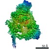


 PDBj
PDBj


 Assembly

 Mass: 94.971 Da / Num. of mol.: 1 / Source method: obtained synthetically / Formula: PO4 / Feature type: SUBJECT OF INVESTIGATION
 Mass: 18.015 Da / Num. of mol.: 109 / Source method: isolated from a natural source / Formula: H2O
 Sample preparation
 Processing