 Movie
Movie Controller


+ Open data

- Basic information
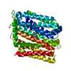
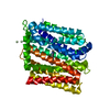
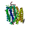
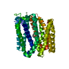
| Entry | Database: PDB / ID: 6vs0 | ||||||
|---|---|---|---|---|---|---|---|
| Title | protein B | ||||||
 Components Components | Multidrug transporter MdfA | ||||||
 Keywords Keywords | TRANSPORT PROTEIN/ANTIBIOTIC / membrane protein / TRANSPORT PROTEIN-ANTIBIOTIC complex | ||||||
| Function / homology |  Function and homology information Function and homology informationpotassium:proton antiporter activity / sodium:proton antiporter activity / xenobiotic detoxification by transmembrane export across the plasma membrane / : / Antimicrobial resistance / response to antibiotic / plasma membrane Similarity search - Function | ||||||
| Biological species |  | ||||||
| Method |  X-RAY DIFFRACTION / SYNCHROTRON / SAD / Resolution: 2.1 Å X-RAY DIFFRACTION / SYNCHROTRON / SAD / Resolution: 2.1 Å | ||||||
 Authors Authors | Lu, M. / Lu, M.M. | ||||||
| Funding support |  United States, 1items United States, 1items
| ||||||
 Citation Citation | Journal: Sci Rep / Year: 2020 Title: Structure and mechanism of a redesigned multidrug transporter from the Major Facilitator Superfamily. Authors: Wu, H.H. / Symersky, J. / Lu, M. | ||||||
| History |
|
- Structure visualization
| Structure viewer | Molecule: MolmilJmol/JSmol |
|---|
- Downloads & links
-Download
| PDBx/mmCIF format | 6vs0.cif.gz | 88.9 KB | Display | PDBx/mmCIF format |
|---|---|---|---|---|
| PDB format | pdb6vs0.ent.gz | 65 KB | Display | PDB format |
| PDBx/mmJSON format | 6vs0.json.gz | Tree view | PDBx/mmJSON format | |
| Others |  Other downloads Other downloads |
-Validation report
| Arichive directory | https://data.pdbj.org/pub/pdb/validation_reports/vs/6vs0ftp://data.pdbj.org/pub/pdb/validation_reports/vs/6vs0 | HTTPS FTP |
|---|
-Related structure data


-Links
 PDBj
PDBj


- Assembly
| Deposited unit | 
| ||||||||
|---|---|---|---|---|---|---|---|---|---|
| 1 |
| ||||||||
| Unit cell |
|
-Components
| #1: Protein | Mass: 41923.020 Da / Num. of mol.: 1 / Mutation: E26T/D34M/A150E Source method: isolated from a genetically manipulated source Source: (gene. exp.) | ||||
|---|---|---|---|---|---|
| #2: Chemical | ChemComp-CLM /   Mass: 323.129 Da / Num. of mol.: 1 / Source method: obtained synthetically / Formula: C11H12Cl2N2O5 / Feature type: SUBJECT OF INVESTIGATION / Comment: antibiotic*YM Mass: 323.129 Da / Num. of mol.: 1 / Source method: obtained synthetically / Formula: C11H12Cl2N2O5 / Feature type: SUBJECT OF INVESTIGATION / Comment: antibiotic*YM | ||||
| #3: Chemical |   Mass: 140.908 Da / Num. of mol.: 2 / Source method: obtained synthetically / Formula: Pr Mass: 140.908 Da / Num. of mol.: 2 / Source method: obtained synthetically / Formula: Pr#4: Water | ChemComp-HOH / |  Mass: 18.015 Da / Num. of mol.: 80 / Source method: isolated from a natural source / Formula: H2O Mass: 18.015 Da / Num. of mol.: 80 / Source method: isolated from a natural source / Formula: H2OHas ligand of interest | Y | |
-Experimental details
-Experiment
| Experiment | Method: X-RAY DIFFRACTION / Number of used crystals: 1 |
|---|
- Sample preparation
| Crystal | Density Matthews: 3.75 Å3/Da / Density % sol: 67.18 % |
|---|---|
| Crystal grow | Temperature: 293 K / Method: vapor diffusion, hanging drop / Details: PEG, salts, etc |
-Data collection
| Diffraction | Mean temperature: 100 K / Serial crystal experiment: N |
|---|---|
| Diffraction source | Source: SYNCHROTRON / Site: APS / Beamline: 23-ID-B / Wavelength: 1 Å |
| Detector | Type: MAR CCD 130 mm / Detector: CCD / Date: Jan 1, 2018 |
| Radiation | Protocol: SINGLE WAVELENGTH / Monochromatic (M) / Laue (L): M / Scattering type: x-ray |
| Radiation wavelength | Wavelength: 1 Å / Relative weight: 1 |
| Reflection | Resolution: 2.1→100 Å / Num. obs: 34557 / % possible obs: 99 % / Redundancy: 11 % / CC1/2: 1 / Rsym value: 0.1 / Net I/σ(I): 31 |
| Reflection shell | Resolution: 2.1→2.2 Å / Num. unique obs: 300 / CC1/2: 0.4 / Rsym value: 0.7 / % possible all: 94 |
- Processing
| Software |
| ||||||||||||||||||
|---|---|---|---|---|---|---|---|---|---|---|---|---|---|---|---|---|---|---|---|
| Refinement | Method to determine structure: SAD / Resolution: 2.1→15 Å / Cross valid method: THROUGHOUT
| ||||||||||||||||||
| Displacement parameters | Biso max: 131.93 Å2 / Biso mean: 63.1509 Å2 / Biso min: 29.47 Å2 | ||||||||||||||||||
| Refinement step | Cycle: LAST / Resolution: 2.1→15 Å
|
