 Movie
Movie Controller

[English] 日本語
 Yorodumi
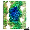
- PDB-6vpp: Cryo-EM structure of microtubule-bound KLP61F motor with tail dom... -
+ Open data

- Basic information
| Entry | Database: PDB / ID: 6vpp | |||||||||
|---|---|---|---|---|---|---|---|---|---|---|
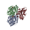
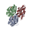
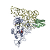
| Title | Cryo-EM structure of microtubule-bound KLP61F motor with tail domain in the nucleotide-free state | |||||||||
 Components Components |
| |||||||||
 Keywords Keywords | MOTOR PROTEIN / Kinesin-5 / KLP61F / microtubules / mitotic spindles / nucleotide-free state / cell cycle | |||||||||
| Function / homology |  Function and homology information Function and homology informationaster / plus-end directed microtubule sliding / fusome organization / fusome / COPI-dependent Golgi-to-ER retrograde traffic / Kinesins / centrosome separation / spindle elongation / mitotic spindle microtubule / mitotic centrosome separation ...aster / plus-end directed microtubule sliding / fusome organization / fusome / COPI-dependent Golgi-to-ER retrograde traffic / Kinesins / centrosome separation / spindle elongation / mitotic spindle microtubule / mitotic centrosome separation / microtubule bundle formation / plus-end-directed microtubule motor activity / microtubule associated complex / positive regulation of Golgi to plasma membrane protein transport / kinesin complex / microtubule-based movement / mitotic spindle pole / Golgi organization / cytoskeletal motor activity / protein secretion / mitotic spindle assembly / mitotic spindle organization / spindle microtubule / microtubule cytoskeleton organization / structural constituent of cytoskeleton / neuron migration / spindle / mitotic spindle / mitotic cell cycle / microtubule binding / microtubule / Hydrolases; Acting on acid anhydrides; Acting on GTP to facilitate cellular and subcellular movement / cell division / hydrolase activity / GTPase activity / GTP binding / Golgi apparatus / endoplasmic reticulum / ATP binding / metal ion binding / nucleus / cytoplasm Similarity search - Function | |||||||||
| Biological species |   | |||||||||
| Method | ELECTRON MICROSCOPY / helical reconstruction / cryo EM / Resolution: 4.4 Å | |||||||||
 Authors Authors | Bodrug, T. / Wilson-Kubalek, E.M. / Nithianantham, S. / Debs, G. / Sindelar, C.V. / Milligan, R. / Al-Bassam, J. | |||||||||
| Funding support |  United States, 2items United States, 2items
| |||||||||
 Citation Citation | Journal: Elife / Year: 2020 Title: The kinesin-5 tail domain directly modulates the mechanochemical cycle of the motor domain for anti-parallel microtubule sliding. Authors: Tatyana Bodrug / Elizabeth M Wilson-Kubalek / Stanley Nithianantham / Alex F Thompson / April Alfieri / Ignas Gaska / Jennifer Major / Garrett Debs / Sayaka Inagaki / Pedro Gutierrez / ...Authors: Tatyana Bodrug / Elizabeth M Wilson-Kubalek / Stanley Nithianantham / Alex F Thompson / April Alfieri / Ignas Gaska / Jennifer Major / Garrett Debs / Sayaka Inagaki / Pedro Gutierrez / Larisa Gheber / Richard J McKenney / Charles Vaughn Sindelar / Ronald Milligan / Jason Stumpff / Steven S Rosenfeld / Scott T Forth / Jawdat Al-Bassam /  Abstract: Kinesin-5 motors organize mitotic spindles by sliding apart microtubules. They are homotetramers with dimeric motor and tail domains at both ends of a bipolar minifilament. Here, we describe a ...Kinesin-5 motors organize mitotic spindles by sliding apart microtubules. They are homotetramers with dimeric motor and tail domains at both ends of a bipolar minifilament. Here, we describe a regulatory mechanism involving direct binding between tail and motor domains and its fundamental role in microtubule sliding. Kinesin-5 tails decrease microtubule-stimulated ATP-hydrolysis by specifically engaging motor domains in the nucleotide-free or ADP states. Cryo-EM reveals that tail binding stabilizes an open motor domain ATP-active site. Full-length motors undergo slow motility and cluster together along microtubules, while tail-deleted motors exhibit rapid motility without clustering. The tail is critical for motors to zipper together two microtubules by generating substantial sliding forces. The tail is essential for mitotic spindle localization, which becomes severely reduced in tail-deleted motors. Our studies suggest a revised microtubule-sliding model, in which kinesin-5 tails stabilize motor domains in the microtubule-bound state by slowing ATP-binding, resulting in high-force production at both homotetramer ends. | |||||||||
| History |
|
- Structure visualization
| Movie |
Movie viewer |
|---|---|
| Structure viewer | Molecule: MolmilJmol/JSmol |
- Downloads & links
-Download
| PDBx/mmCIF format | 6vpp.cif.gz | 219.6 KB | Display | PDBx/mmCIF format |
|---|---|---|---|---|
| PDB format | pdb6vpp.ent.gz | 169.5 KB | Display | PDB format |
| PDBx/mmJSON format | 6vpp.json.gz | Tree view | PDBx/mmJSON format | |
| Others |  Other downloads Other downloads |
-Validation report
| Arichive directory | https://data.pdbj.org/pub/pdb/validation_reports/vp/6vppftp://data.pdbj.org/pub/pdb/validation_reports/vp/6vpp | HTTPS FTP |
|---|
-Related structure data
| Related structure data | 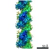 21315MC 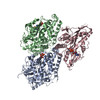 6vpoC M: map data used to model this data C: citing same article ( |
|---|---|
| Similar structure data |


-Links
 PDBj
PDBj


- Assembly
| Deposited unit | 
|
|---|---|
| 1 |
|
-Components
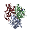
-Protein , 3 types, 3 molecules ABC
| #1: Protein | Mass: 50121.266 Da / Num. of mol.: 1 / Source method: isolated from a natural source / Source: (natural) |
|---|---|
| #2: Protein | Mass: 49907.770 Da / Num. of mol.: 1 / Source method: isolated from a natural source / Source: (natural) |
| #3: Protein | Mass: 42627.336 Da / Num. of mol.: 1 Source method: isolated from a genetically manipulated source Source: (gene. exp.)  |
-Non-polymers , 3 types, 3 molecules 


| #4: Chemical | ChemComp-GTP /  Mass: 523.180 Da / Num. of mol.: 1 / Source method: obtained synthetically / Formula: C10H16N5O14P3 / Comment: GTP, energy-carrying molecule*YM Mass: 523.180 Da / Num. of mol.: 1 / Source method: obtained synthetically / Formula: C10H16N5O14P3 / Comment: GTP, energy-carrying molecule*YM |
|---|---|
| #5: Chemical | ChemComp-MG /  Mass: 24.305 Da / Num. of mol.: 1 / Source method: obtained synthetically / Formula: Mg Mass: 24.305 Da / Num. of mol.: 1 / Source method: obtained synthetically / Formula: Mg |
| #6: Chemical | ChemComp-GDP /  Type: RNA linking / Mass: 443.201 Da / Num. of mol.: 1 / Source method: obtained synthetically / Formula: C10H15N5O11P2 / Feature type: SUBJECT OF INVESTIGATION / Comment: GDP, energy-carrying molecule*YM Type: RNA linking / Mass: 443.201 Da / Num. of mol.: 1 / Source method: obtained synthetically / Formula: C10H15N5O11P2 / Feature type: SUBJECT OF INVESTIGATION / Comment: GDP, energy-carrying molecule*YM |
-Details
| Has ligand of interest | Y |
|---|
-Experimental details
-Experiment
| Experiment | Method: ELECTRON MICROSCOPY |
|---|---|
| EM experiment | Aggregation state: HELICAL ARRAY / 3D reconstruction method: helical reconstruction |
- Sample preparation
| Component |
| ||||||||||||||||||||||||
|---|---|---|---|---|---|---|---|---|---|---|---|---|---|---|---|---|---|---|---|---|---|---|---|---|---|
| Molecular weight | Units: KILODALTONS/NANOMETER / Experimental value: YES | ||||||||||||||||||||||||
| Source (natural) |
| ||||||||||||||||||||||||
| Source (recombinant) | Organism: | ||||||||||||||||||||||||
| Buffer solution | pH: 6.8 | ||||||||||||||||||||||||
| Specimen | Embedding applied: NO / Shadowing applied: NO / Staining applied: NO / Vitrification applied: YES | ||||||||||||||||||||||||
| Specimen support | Grid material: GOLD / Grid mesh size: 400 divisions/in. / Grid type: Quantifoil, UltrAuFoil, R1.2/1.3 | ||||||||||||||||||||||||
| Vitrification | Instrument: HOMEMADE PLUNGER / Cryogen name: ETHANE / Humidity: 90 % / Chamber temperature: 298 K |
- Electron microscopy imaging
| Experimental equipment |  Model: Titan Krios / Image courtesy: FEI Company |
|---|---|
| Microscopy | Model: FEI TITAN KRIOS |
| Electron gun | Electron source:  FIELD EMISSION GUN / Accelerating voltage: 300 kV / Illumination mode: FLOOD BEAM FIELD EMISSION GUN / Accelerating voltage: 300 kV / Illumination mode: FLOOD BEAM |
| Electron lens | Mode: BRIGHT FIELD |
| Image recording | Electron dose: 38 e/Å2 / Film or detector model: GATAN K2 SUMMIT (4k x 4k) |
- Processing
| EM software |
| ||||||||||||||||||||||||
|---|---|---|---|---|---|---|---|---|---|---|---|---|---|---|---|---|---|---|---|---|---|---|---|---|---|
| CTF correction | Type: PHASE FLIPPING AND AMPLITUDE CORRECTION | ||||||||||||||||||||||||
| Helical symmerty | Angular rotation/subunit: -34.61 ° / Axial rise/subunit: 17.4 Å / Axial symmetry: C1 | ||||||||||||||||||||||||
| 3D reconstruction | Resolution: 4.4 Å / Resolution method: FSC 0.143 CUT-OFF / Num. of particles: 14570 / Symmetry type: HELICAL | ||||||||||||||||||||||||
| Atomic model building | B value: 208 / Protocol: FLEXIBLE FIT / Space: REAL / Target criteria: Correlation coefficient |
