+ Open data
- Basic information
| Entry | Database: PDB / ID: 6voy | ||||||||||||||||||||||||
|---|---|---|---|---|---|---|---|---|---|---|---|---|---|---|---|---|---|---|---|---|---|---|---|---|---|
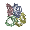
| Title | Cryo-EM structure of HTLV-1 instasome | ||||||||||||||||||||||||
 Components Components |
| ||||||||||||||||||||||||
 Keywords Keywords | DNA BINDING PROTEIN/DNA / Integrase / Intasome / DNA BINDING PROTEIN-DNA complex | ||||||||||||||||||||||||
| Function / homology |  Function and homology information Function and homology informationprotein phosphatase type 2A complex / meiotic sister chromatid cohesion / protein phosphatase regulator activity / APC truncation mutants have impaired AXIN binding / AXIN missense mutants destabilize the destruction complex / Truncations of AMER1 destabilize the destruction complex / Beta-catenin phosphorylation cascade / Signaling by GSK3beta mutants / CTNNB1 S33 mutants aren't phosphorylated / CTNNB1 S37 mutants aren't phosphorylated ...protein phosphatase type 2A complex / meiotic sister chromatid cohesion / protein phosphatase regulator activity / APC truncation mutants have impaired AXIN binding / AXIN missense mutants destabilize the destruction complex / Truncations of AMER1 destabilize the destruction complex / Beta-catenin phosphorylation cascade / Signaling by GSK3beta mutants / CTNNB1 S33 mutants aren't phosphorylated / CTNNB1 S37 mutants aren't phosphorylated / CTNNB1 S45 mutants aren't phosphorylated / CTNNB1 T41 mutants aren't phosphorylated / Co-stimulation by CD28 / Disassembly of the destruction complex and recruitment of AXIN to the membrane / Co-inhibition by CTLA4 / Platelet sensitization by LDL / protein phosphatase activator activity / chromosome, centromeric region / intrinsic apoptotic signaling pathway in response to DNA damage by p53 class mediator / Amplification of signal from unattached kinetochores via a MAD2 inhibitory signal / Mitotic Prometaphase / EML4 and NUDC in mitotic spindle formation / RNA endonuclease activity / Resolution of Sister Chromatid Cohesion / DNA damage response, signal transduction by p53 class mediator / RHO GTPases Activate Formins / RAF activation / Degradation of beta-catenin by the destruction complex / DNA integration / viral genome integration into host DNA / establishment of integrated proviral latency / RNA stem-loop binding / RNA-directed DNA polymerase activity / RNA-DNA hybrid ribonuclease activity / Negative regulation of MAPK pathway / Separation of Sister Chromatids / Regulation of TP53 Degradation / PI5P, PP2A and IER3 Regulate PI3K/AKT Signaling / DNA recombination / proteasome-mediated ubiquitin-dependent protein catabolic process / negative regulation of cell population proliferation / symbiont entry into host cell / Golgi apparatus / signal transduction / DNA binding / zinc ion binding / nucleoplasm / nucleus / cytoplasm / cytosol Similarity search - Function | ||||||||||||||||||||||||
| Biological species |   Saccharolobus solfataricus (archaea) Saccharolobus solfataricus (archaea) Human T-cell leukemia virus type I Human T-cell leukemia virus type I Homo sapiens (human) Homo sapiens (human) | ||||||||||||||||||||||||
| Method | ELECTRON MICROSCOPY / single particle reconstruction / cryo EM / Resolution: 3.7 Å | ||||||||||||||||||||||||
 Authors Authors | Bhatt, V. / Shi, K. / Sundborger, A. / Aihara, H. | ||||||||||||||||||||||||
| Funding support |  United States, 1items United States, 1items
| ||||||||||||||||||||||||
 Citation Citation |  Journal: Nat Commun / Year: 2020 Journal: Nat Commun / Year: 2020Title: Structural basis of host protein hijacking in human T-cell leukemia virus integration. Authors: Veer Bhatt / Ke Shi / Daniel J Salamango / Nicholas H Moeller / Krishan K Pandey / Sibes Bera / Heather O Bohl / Fredy Kurniawan / Kayo Orellana / Wei Zhang / Duane P Grandgenett / Reuben S ...Authors: Veer Bhatt / Ke Shi / Daniel J Salamango / Nicholas H Moeller / Krishan K Pandey / Sibes Bera / Heather O Bohl / Fredy Kurniawan / Kayo Orellana / Wei Zhang / Duane P Grandgenett / Reuben S Harris / Anna C Sundborger-Lunna / Hideki Aihara /  Abstract: Integration of the reverse-transcribed viral DNA into host chromosomes is a critical step in the life-cycle of retroviruses, including an oncogenic delta(δ)-retrovirus human T-cell leukemia virus ...Integration of the reverse-transcribed viral DNA into host chromosomes is a critical step in the life-cycle of retroviruses, including an oncogenic delta(δ)-retrovirus human T-cell leukemia virus type-1 (HTLV-1). Retroviral integrase forms a higher order nucleoprotein assembly (intasome) to catalyze the integration reaction, in which the roles of host factors remain poorly understood. Here, we use cryo-electron microscopy to visualize the HTLV-1 intasome at 3.7-Å resolution. The structure together with functional analyses reveal that the B56γ (B'γ) subunit of an essential host enzyme, protein phosphatase 2 A (PP2A), is repurposed as an integral component of the intasome to mediate HTLV-1 integration. Our studies reveal a key host-virus interaction underlying the replication of an important human pathogen and highlight divergent integration strategies of retroviruses. | ||||||||||||||||||||||||
| History |
|
- Structure visualization
| Movie |
 Movie viewer Movie viewer |
|---|---|
| Structure viewer | Molecule:  Molmil Molmil Jmol/JSmol Jmol/JSmol |
- Downloads & links
- Download
| PDBx/mmCIF format |  6voy.cif.gz 6voy.cif.gz | 413.7 KB | Display |  PDBx/mmCIF format PDBx/mmCIF format |
|---|---|---|---|---|
| PDB format |  pdb6voy.ent.gz pdb6voy.ent.gz | 318.8 KB | Display |  PDB format PDB format |
| PDBx/mmJSON format |  6voy.json.gz 6voy.json.gz | Tree view |  PDBx/mmJSON format PDBx/mmJSON format | |
| Others |  Other downloads Other downloads |
-Validation report
| Arichive directory |  https://data.pdbj.org/pub/pdb/validation_reports/vo/6voy https://data.pdbj.org/pub/pdb/validation_reports/vo/6voy ftp://data.pdbj.org/pub/pdb/validation_reports/vo/6voy ftp://data.pdbj.org/pub/pdb/validation_reports/vo/6voy | HTTPS FTP |
|---|
-Related structure data
| Related structure data |  21301MC M: map data used to model this data C: citing same article ( |
|---|---|
| Similar structure data |
- Links
Links
- Assembly
| Deposited unit | 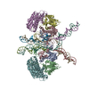
|
|---|---|
| 1 |
|
- Components
-Protein , 2 types, 6 molecules ABCDEF
| #1: Protein | Mass: 43652.680 Da / Num. of mol.: 4 / Mutation: W24A Source method: isolated from a genetically manipulated source Source: (gene. exp.)   Saccharolobus solfataricus (strain ATCC 35092 / DSM 1617 / JCM 11322 / P2) (archaea), (gene. exp.) Saccharolobus solfataricus (strain ATCC 35092 / DSM 1617 / JCM 11322 / P2) (archaea), (gene. exp.)  Human T-cell leukemia virus type I Human T-cell leukemia virus type IStrain: ATCC 35092 / DSM 1617 / JCM 11322 / P2 / Gene: sso7d, sso7d-1, SSO10610, pol / Production host:  #2: Protein | Mass: 40357.789 Da / Num. of mol.: 2 Source method: isolated from a genetically manipulated source Source: (gene. exp.)  Homo sapiens (human) / Gene: PPP2R5C, KIAA0044 / Production host: Homo sapiens (human) / Gene: PPP2R5C, KIAA0044 / Production host:  |
|---|
-DNA (5'-D(P*AP*CP*AP*CP*AP*CP*TP*TP*GP*AP*CP*TP*AP*GP*GP*GP*TP*G)- ... , 2 types, 4 molecules ILKN
| #3: DNA chain | Mass: 15074.711 Da / Num. of mol.: 2 / Source method: obtained synthetically / Source: (synth.)  Homo sapiens (human) Homo sapiens (human)#5: DNA chain | Mass: 6199.017 Da / Num. of mol.: 2 / Source method: obtained synthetically / Source: (synth.)  Human T-cell leukemia virus type I Human T-cell leukemia virus type I |
|---|
-DNA chain , 1 types, 2 molecules JM
| #4: DNA chain | Mass: 7614.918 Da / Num. of mol.: 2 / Source method: obtained synthetically / Source: (synth.)  Human T-cell leukemia virus type I Human T-cell leukemia virus type I |
|---|
-Non-polymers , 2 types, 6 molecules 


| #6: Chemical | ChemComp-ZN / #7: Chemical | |
|---|
-Details
| Has ligand of interest | N |
|---|---|
| Has protein modification | Y |
-Experimental details
-Experiment
| Experiment | Method: ELECTRON MICROSCOPY |
|---|---|
| EM experiment | Aggregation state: PARTICLE / 3D reconstruction method: single particle reconstruction |
- Sample preparation
| Component | Name: HTLV1 intasome / Type: COMPLEX / Entity ID: #1-#5 / Source: RECOMBINANT |
|---|---|
| Source (natural) | Organism:  Human T-cell leukemia virus type I Human T-cell leukemia virus type I |
| Source (recombinant) | Organism:  |
| Buffer solution | pH: 7.5 |
| Specimen | Embedding applied: NO / Shadowing applied: NO / Staining applied: NO / Vitrification applied: YES |
| Vitrification | Cryogen name: NITROGEN |
- Electron microscopy imaging
| Experimental equipment |  Model: Titan Krios / Image courtesy: FEI Company |
|---|---|
| Microscopy | Model: FEI TITAN KRIOS |
| Electron gun | Electron source:  FIELD EMISSION GUN / Accelerating voltage: 300 kV / Illumination mode: FLOOD BEAM FIELD EMISSION GUN / Accelerating voltage: 300 kV / Illumination mode: FLOOD BEAM |
| Electron lens | Mode: BRIGHT FIELD |
| Image recording | Electron dose: 30 e/Å2 / Film or detector model: FEI FALCON III (4k x 4k) |
- Processing
| Software |
| ||||||||||||||||||||||||
|---|---|---|---|---|---|---|---|---|---|---|---|---|---|---|---|---|---|---|---|---|---|---|---|---|---|
| EM software |
| ||||||||||||||||||||||||
| CTF correction | Type: PHASE FLIPPING AND AMPLITUDE CORRECTION | ||||||||||||||||||||||||
| Symmetry | Point symmetry: C2 (2 fold cyclic) | ||||||||||||||||||||||||
| 3D reconstruction | Resolution: 3.7 Å / Resolution method: FSC 0.143 CUT-OFF / Num. of particles: 30434 / Symmetry type: POINT | ||||||||||||||||||||||||
| Refinement | Cross valid method: NONE Stereochemistry target values: GeoStd + Monomer Library + CDL v1.2 | ||||||||||||||||||||||||
| Displacement parameters | Biso mean: 189.65 Å2 | ||||||||||||||||||||||||
| Refine LS restraints |
|
 Movie
Movie Controller


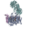

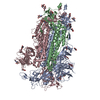
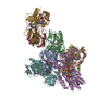


 PDBj
PDBj


