 Movie
Movie Controller


+ Open data

- Basic information
| Entry | Database: EMDB / ID: EMD-21301 | |||||||||
|---|---|---|---|---|---|---|---|---|---|---|
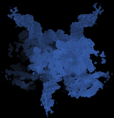
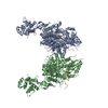
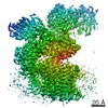
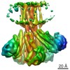
| Title | Cryo-EM structure of HTLV-1 instasome | |||||||||
 Map data Map data | Intasome | |||||||||
 Sample Sample |
| |||||||||
 Keywords Keywords | Integrase / Intasome / DNA BINDING PROTEIN-DNA complex | |||||||||
| Function / homology |  Function and homology information Function and homology informationprotein phosphatase type 2A complex / meiotic sister chromatid cohesion / protein phosphatase regulator activity / APC truncation mutants have impaired AXIN binding / AXIN missense mutants destabilize the destruction complex / Truncations of AMER1 destabilize the destruction complex / Beta-catenin phosphorylation cascade / Signaling by GSK3beta mutants / CTNNB1 S33 mutants aren't phosphorylated / CTNNB1 S37 mutants aren't phosphorylated ...protein phosphatase type 2A complex / meiotic sister chromatid cohesion / protein phosphatase regulator activity / APC truncation mutants have impaired AXIN binding / AXIN missense mutants destabilize the destruction complex / Truncations of AMER1 destabilize the destruction complex / Beta-catenin phosphorylation cascade / Signaling by GSK3beta mutants / CTNNB1 S33 mutants aren't phosphorylated / CTNNB1 S37 mutants aren't phosphorylated / CTNNB1 S45 mutants aren't phosphorylated / CTNNB1 T41 mutants aren't phosphorylated / Co-stimulation by CD28 / Disassembly of the destruction complex and recruitment of AXIN to the membrane / Co-inhibition by CTLA4 / Platelet sensitization by LDL / protein phosphatase activator activity / intrinsic apoptotic signaling pathway in response to DNA damage by p53 class mediator / chromosome, centromeric region / RNA endonuclease activity / Amplification of signal from unattached kinetochores via a MAD2 inhibitory signal / Mitotic Prometaphase / EML4 and NUDC in mitotic spindle formation / Resolution of Sister Chromatid Cohesion / DNA damage response, signal transduction by p53 class mediator / RAF activation / DNA integration / RHO GTPases Activate Formins / viral genome integration into host DNA / establishment of integrated proviral latency / RNA stem-loop binding / Degradation of beta-catenin by the destruction complex / RNA-directed DNA polymerase activity / RNA-DNA hybrid ribonuclease activity / Negative regulation of MAPK pathway / Separation of Sister Chromatids / Regulation of TP53 Degradation / PI5P, PP2A and IER3 Regulate PI3K/AKT Signaling / DNA recombination / proteasome-mediated ubiquitin-dependent protein catabolic process / negative regulation of cell population proliferation / symbiont entry into host cell / Golgi apparatus / signal transduction / DNA binding / zinc ion binding / nucleoplasm / nucleus / cytoplasm / cytosol Similarity search - Function | |||||||||
| Biological species |  Human T-cell leukemia virus type I / Human T-cell leukemia virus type I /  Homo sapiens (human) Homo sapiens (human) | |||||||||
| Method | single particle reconstruction / cryo EM / Resolution: 3.7 Å | |||||||||
 Authors Authors | Bhatt V / Shi K / Sundborger A / Aihara H | |||||||||
| Funding support |  United States, 1 items United States, 1 items
| |||||||||
 Citation Citation | Journal: Nat Commun / Year: 2020 Title: Structural basis of host protein hijacking in human T-cell leukemia virus integration. Authors: Veer Bhatt / Ke Shi / Daniel J Salamango / Nicholas H Moeller / Krishan K Pandey / Sibes Bera / Heather O Bohl / Fredy Kurniawan / Kayo Orellana / Wei Zhang / Duane P Grandgenett / Reuben S ...Authors: Veer Bhatt / Ke Shi / Daniel J Salamango / Nicholas H Moeller / Krishan K Pandey / Sibes Bera / Heather O Bohl / Fredy Kurniawan / Kayo Orellana / Wei Zhang / Duane P Grandgenett / Reuben S Harris / Anna C Sundborger-Lunna / Hideki Aihara / Abstract: Integration of the reverse-transcribed viral DNA into host chromosomes is a critical step in the life-cycle of retroviruses, including an oncogenic delta(δ)-retrovirus human T-cell leukemia virus ...Integration of the reverse-transcribed viral DNA into host chromosomes is a critical step in the life-cycle of retroviruses, including an oncogenic delta(δ)-retrovirus human T-cell leukemia virus type-1 (HTLV-1). Retroviral integrase forms a higher order nucleoprotein assembly (intasome) to catalyze the integration reaction, in which the roles of host factors remain poorly understood. Here, we use cryo-electron microscopy to visualize the HTLV-1 intasome at 3.7-Å resolution. The structure together with functional analyses reveal that the B56γ (B'γ) subunit of an essential host enzyme, protein phosphatase 2 A (PP2A), is repurposed as an integral component of the intasome to mediate HTLV-1 integration. Our studies reveal a key host-virus interaction underlying the replication of an important human pathogen and highlight divergent integration strategies of retroviruses. | |||||||||
| History |
|
- Structure visualization
| Movie |
Movie viewer |
|---|---|
| Structure viewer | EM map: SurfViewMolmilJmol/JSmol |
| Supplemental images |
- Downloads & links
-EMDB archive
| Map data | emd_21301.map.gz | 65.7 MB | EMDB map data format | |
|---|---|---|---|---|
| Header (meta data) | emd-21301-v30.xmlemd-21301.xml | 16.9 KB 16.9 KB | Display Display | EMDB header |
| Images |  emd_21301.png emd_21301.png | 130.3 KB | ||
| Filedesc metadata | emd-21301.cif.gz | 6.9 KB | ||
| Archive directory |  http://ftp.pdbj.org/pub/emdb/structures/EMD-21301ftp://ftp.pdbj.org/pub/emdb/structures/EMD-21301 http://ftp.pdbj.org/pub/emdb/structures/EMD-21301ftp://ftp.pdbj.org/pub/emdb/structures/EMD-21301 | HTTPS FTP |
-Related structure data
| Related structure data |  6voyMC M: atomic model generated by this map C: citing same article ( |
|---|---|
| Similar structure data |


-Links
| EMDB pages | EMDB (EBI/PDBe) / EMDataResource |
|---|---|
| Related items in Molecule of the Month |


-Map
| File | Download / File: emd_21301.map.gz / Format: CCP4 / Size: 70.2 MB / Type: IMAGE STORED AS FLOATING POINT NUMBER (4 BYTES) | ||||||||||||||||||||||||||||||||||||||||||||||||||||||||||||||||||||
|---|---|---|---|---|---|---|---|---|---|---|---|---|---|---|---|---|---|---|---|---|---|---|---|---|---|---|---|---|---|---|---|---|---|---|---|---|---|---|---|---|---|---|---|---|---|---|---|---|---|---|---|---|---|---|---|---|---|---|---|---|---|---|---|---|---|---|---|---|---|
| Annotation | Intasome | ||||||||||||||||||||||||||||||||||||||||||||||||||||||||||||||||||||
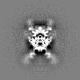
| Projections & slices | Image control
Images are generated by Spider. | ||||||||||||||||||||||||||||||||||||||||||||||||||||||||||||||||||||
| Voxel size | X=Y=Z: 0.8933 Å | ||||||||||||||||||||||||||||||||||||||||||||||||||||||||||||||||||||
| Density |
| ||||||||||||||||||||||||||||||||||||||||||||||||||||||||||||||||||||
| Symmetry | Space group: 1 | ||||||||||||||||||||||||||||||||||||||||||||||||||||||||||||||||||||
| Details | EMDB XML:
CCP4 map header:
| ||||||||||||||||||||||||||||||||||||||||||||||||||||||||||||||||||||
 Z (Sec.)
 Y (Row.)
 X (Col.)


-Supplemental data
- Sample components
-Entire : HTLV1 intasome
| Entire | Name: HTLV1 intasome |
|---|---|
| Components |
|
-Supramolecule #1: HTLV1 intasome
| Supramolecule | Name: HTLV1 intasome / type: complex / ID: 1 / Parent: 0 / Macromolecule list: #1-#5 |
|---|---|
| Source (natural) | Organism: Human T-cell leukemia virus type I |
-Macromolecule #1: DNA-binding protein 7d
| Macromolecule | Name: DNA-binding protein 7d / type: protein_or_peptide / ID: 1 / Number of copies: 4 / Enantiomer: LEVO |
|---|---|
| Source (natural) | Organism: Human T-cell leukemia virus type I |
| Molecular weight | Theoretical: 43.65268 KDa |
| Recombinant expression | Organism:  |
| Sequence | String: MGSSHHHHHH SSGLVPRGSH MATVKFKYKG EEKEVDISKI KKVARVGKMI SFTYDEGGGK TGEGAVSEKD APKELLQMLE KQKKGGSLE VLFQGPSPAE LHSFTHCGQT ALTLQGATTT EASNILRSCH ACRKNNPQHQ MPRGHIRRGL LPNHIWQGDI T HFKYKNTL ...String: MGSSHHHHHH SSGLVPRGSH MATVKFKYKG EEKEVDISKI KKVARVGKMI SFTYDEGGGK TGEGAVSEKD APKELLQMLE KQKKGGSLE VLFQGPSPAE LHSFTHCGQT ALTLQGATTT EASNILRSCH ACRKNNPQHQ MPRGHIRRGL LPNHIWQGDI T HFKYKNTL YRLHVWVDTF SGAISATQKR KETSSEAISS LLQAIAYLGK PSYINTDNGP AYISQDFLNM CTSLAIRHTT HV PYNPTSS GLVQRSNGIL KTLLYKYFTD KPDLPMDNAL SIALWTINHL NVLTNCHKTR WQLHHSPRLQ PIPETRSLSN KQT HWYYFK LPGLNSRQWK GPQEALQEAA GAALIPVSAS SAQWIPWRLL KRAACPRPVG GPADPKEKDH QHHG UniProtKB: DNA-binding protein 7d, Pol protein |
-Macromolecule #2: Serine/threonine-protein phosphatase 2A 56 kDa regulatory subunit...
| Macromolecule | Name: Serine/threonine-protein phosphatase 2A 56 kDa regulatory subunit gamma isoform type: protein_or_peptide / ID: 2 / Number of copies: 2 / Enantiomer: LEVO |
|---|---|
| Source (natural) | Organism: Homo sapiens (human) |
| Molecular weight | Theoretical: 40.357789 KDa |
| Recombinant expression | Organism: |
| Sequence | String: IRDVPPADQE KLFIQKLRQC CVLFDFVSDP LSDLKWKEVK RAALSEMVEY ITHNRNVITE PIYPEVVHMF AVNMFRTLPP SSNPTGAEF DPEEDEPTLE AAWPHLQLVY EFFLRFLESP DFQPNIAKKY IDQKFVLQLL ELFDSEDPRE RDFLKTTLHR I YGKFLGLR ...String: IRDVPPADQE KLFIQKLRQC CVLFDFVSDP LSDLKWKEVK RAALSEMVEY ITHNRNVITE PIYPEVVHMF AVNMFRTLPP SSNPTGAEF DPEEDEPTLE AAWPHLQLVY EFFLRFLESP DFQPNIAKKY IDQKFVLQLL ELFDSEDPRE RDFLKTTLHR I YGKFLGLR AYIRKQINNI FYRFIYETEH HNGIAELLEI LGSIINGFAL PLKEEHKIFL LKVLLPLHKV KSLSVYHPQL AY CVVQFLE KDSTLTEPVV MALLKYWPKT HSPKEVMFLN ELEEILDVIE PSEFVKIMEP LFRQLAKCVS SPHFQVAERA LYY WNNEYI MSLISDNAAK ILPIMFP UniProtKB: Serine/threonine-protein phosphatase 2A 56 kDa regulatory subunit gamma isoform |
-Macromolecule #3: DNA (5'-D(P*AP*CP*AP*CP*AP*CP*TP*TP*GP*AP*CP*TP*AP*GP*GP*GP*TP*G)-3')
| Macromolecule | Name: DNA (5'-D(P*AP*CP*AP*CP*AP*CP*TP*TP*GP*AP*CP*TP*AP*GP*GP*GP*TP*G)-3') type: dna / ID: 3 / Number of copies: 2 / Classification: DNA |
|---|---|
| Source (natural) | Organism: Homo sapiens (human) |
| Molecular weight | Theoretical: 15.074711 KDa |
| Sequence | String: (DC)(DC)(DA)(DG)(DG)(DA)(DG)(DA)(DG)(DA) (DA)(DA)(DT)(DT)(DT)(DA)(DG)(DT)(DA)(DC) (DA)(DC)(DA)(DG)(DA)(DT)(DA)(DT)(DC) (DC)(DA)(DC)(DC)(DC)(DT)(DA)(DG)(DT)(DC) (DA) (DA)(DG)(DT)(DG)(DT)(DG)(DT)(DC) (DC) |
-Macromolecule #4: DNA (25-MER)
| Macromolecule | Name: DNA (25-MER) / type: dna / ID: 4 / Number of copies: 2 / Classification: DNA |
|---|---|
| Source (natural) | Organism: Human T-cell leukemia virus type I |
| Molecular weight | Theoretical: 7.614918 KDa |
| Sequence | String: (DA)(DC)(DT)(DG)(DT)(DG)(DT)(DA)(DC)(DT) (DA)(DA)(DA)(DT)(DT)(DT)(DC)(DT)(DC)(DT) (DC)(DC)(DT)(DG)(DG) |
-Macromolecule #5: DNA (5'-D(P*AP*CP*AP*CP*AP*CP*TP*TP*GP*AP*CP*TP*AP*GP*GP*GP*TP*G)-3')
| Macromolecule | Name: DNA (5'-D(P*AP*CP*AP*CP*AP*CP*TP*TP*GP*AP*CP*TP*AP*GP*GP*GP*TP*G)-3') type: dna / ID: 5 / Number of copies: 2 / Classification: DNA |
|---|---|
| Source (natural) | Organism: Human T-cell leukemia virus type I |
| Molecular weight | Theoretical: 6.199017 KDa |
| Sequence | String: (DG)(DG)(DA)(DC)(DA)(DC)(DA)(DC)(DT)(DT) (DG)(DA)(DC)(DT)(DA)(DG)(DG)(DG)(DT)(DG) |
-Macromolecule #6: ZINC ION
| Macromolecule | Name: ZINC ION / type: ligand / ID: 6 / Number of copies: 4 / Formula: ZN |
|---|---|
| Molecular weight | Theoretical: 65.409 Da |
-Macromolecule #7: MAGNESIUM ION
| Macromolecule | Name: MAGNESIUM ION / type: ligand / ID: 7 / Number of copies: 2 / Formula: MG |
|---|---|
| Molecular weight | Theoretical: 24.305 Da |
-Experimental details
-Structure determination
| Method | cryo EM |
|---|---|
 Processing Processing | single particle reconstruction |
| Aggregation state | particle |
-Sample preparation
| Buffer | pH: 7.5 |
|---|---|
| Vitrification | Cryogen name: NITROGEN |
- Electron microscopy
| Microscope | FEI TITAN KRIOS |
|---|---|
| Image recording | Film or detector model: FEI FALCON III (4k x 4k) / Average electron dose: 30.0 e/Å2 |
| Electron beam | Acceleration voltage: 300 kV / Electron source:  FIELD EMISSION GUN FIELD EMISSION GUN |
| Electron optics | Illumination mode: FLOOD BEAM / Imaging mode: BRIGHT FIELD |
| Experimental equipment |  Model: Titan Krios / Image courtesy: FEI Company |
