 Movie
Movie Controller

[English] 日本語
 Yorodumi
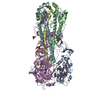
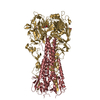
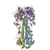
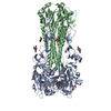
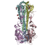
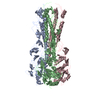
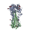
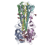
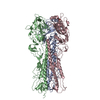
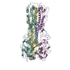
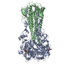
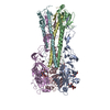
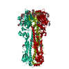
- PDB-6vmz: Crystal Structure of a H5N1 influenza virus hemagglutinin with CBS1117 -
+ Open data

- Basic information
| Entry | Database: PDB / ID: 6vmz | ||||||
|---|---|---|---|---|---|---|---|
| Title | Crystal Structure of a H5N1 influenza virus hemagglutinin with CBS1117 | ||||||
 Components Components | (Hemagglutinin) x 2 | ||||||
 Keywords Keywords | VIRAL PROTEIN / Influenza / H5 / CBS1117 | ||||||
| Function / homology |  Function and homology information Function and homology informationclathrin-dependent endocytosis of virus by host cell / apical plasma membrane / host cell surface receptor binding / fusion of virus membrane with host plasma membrane / fusion of virus membrane with host endosome membrane / viral envelope / virion attachment to host cell / host cell plasma membrane / virion membrane Similarity search - Function | ||||||
| Biological species |   Influenza A virus Influenza A virus | ||||||
| Method |  X-RAY DIFFRACTION / SYNCHROTRON / MOLECULAR REPLACEMENT / molecular replacement / Resolution: 2.2 Å X-RAY DIFFRACTION / SYNCHROTRON / MOLECULAR REPLACEMENT / molecular replacement / Resolution: 2.2 Å | ||||||
 Authors Authors | Antanasijevic, A. / Durst, M.A. / Lavie, A. / Caffrey, M. | ||||||
 Citation Citation | Journal: Life Sci Alliance / Year: 2020 Title: Structure of avian influenza hemagglutinin in complex with a small molecule entry inhibitor. Authors: Antanasijevic, A. / Durst, M.A. / Cheng, H. / Gaisina, I.N. / Perez, J.T. / Manicassamy, B. / Rong, L. / Lavie, A. / Caffrey, M. | ||||||
| History |
|
- Structure visualization
| Structure viewer | Molecule: MolmilJmol/JSmol |
|---|
- Downloads & links
-Download
| PDBx/mmCIF format | 6vmz.cif.gz | 315.2 KB | Display | PDBx/mmCIF format |
|---|---|---|---|---|
| PDB format | pdb6vmz.ent.gz | 252.5 KB | Display | PDB format |
| PDBx/mmJSON format | 6vmz.json.gz | Tree view | PDBx/mmJSON format | |
| Others |  Other downloads Other downloads |
-Validation report
| Arichive directory | https://data.pdbj.org/pub/pdb/validation_reports/vm/6vmzftp://data.pdbj.org/pub/pdb/validation_reports/vm/6vmz | HTTPS FTP |
|---|
-Related structure data
| Related structure data |  2fkoS S: Starting model for refinement |
|---|---|
| Similar structure data |


-Links
 PDBj
PDBj


- Assembly
| Deposited unit | 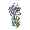
| ||||||||||||||||||||||||||||||||||||||||||||||||||||||||||||||||||||||||||||||||||||||||||||||||||||||||||||||||||||||||||||||||||||||||||||||||||||||
|---|---|---|---|---|---|---|---|---|---|---|---|---|---|---|---|---|---|---|---|---|---|---|---|---|---|---|---|---|---|---|---|---|---|---|---|---|---|---|---|---|---|---|---|---|---|---|---|---|---|---|---|---|---|---|---|---|---|---|---|---|---|---|---|---|---|---|---|---|---|---|---|---|---|---|---|---|---|---|---|---|---|---|---|---|---|---|---|---|---|---|---|---|---|---|---|---|---|---|---|---|---|---|---|---|---|---|---|---|---|---|---|---|---|---|---|---|---|---|---|---|---|---|---|---|---|---|---|---|---|---|---|---|---|---|---|---|---|---|---|---|---|---|---|---|---|---|---|---|---|---|---|
| 1 |
| ||||||||||||||||||||||||||||||||||||||||||||||||||||||||||||||||||||||||||||||||||||||||||||||||||||||||||||||||||||||||||||||||||||||||||||||||||||||
| Unit cell |
| ||||||||||||||||||||||||||||||||||||||||||||||||||||||||||||||||||||||||||||||||||||||||||||||||||||||||||||||||||||||||||||||||||||||||||||||||||||||
| Noncrystallographic symmetry (NCS) | NCS domain:
NCS domain segments: Component-ID: _ / Beg auth comp-ID: GLY / Beg label comp-ID: GLY / Refine code: _
NCS ensembles :
|
-Components
| #1: Protein | Mass: 37948.020 Da / Num. of mol.: 3 / Fragment: N-terminal domain (UNP residues 17-337) Source method: isolated from a genetically manipulated source Source: (gene. exp.) Influenza A virus (A/chicken/Vietnam/4/2003(H5N1))Strain: A/chicken/Vietnam/4/2003(H5N1) / Gene: HA / Production host:  Trichoplusia ni (cabbage looper) / References: UniProt: Q1KHJ8 Trichoplusia ni (cabbage looper) / References: UniProt: Q1KHJ8#2: Protein | Mass: 20881.104 Da / Num. of mol.: 3 / Fragment: C-terminal domain (UNP residues 347-521) Source method: isolated from a genetically manipulated source Source: (gene. exp.) Influenza A virus (A/chicken/Vietnam/30/2003(H5N1))Strain: A/chicken/Vietnam/30/2003(H5N1) / Gene: HA / Production host: Trichoplusia ni (cabbage looper) / References: UniProt: Q1KHK7#3: Sugar | ChemComp-NAG /   Type: D-saccharide, beta linking / Mass: 221.208 Da / Num. of mol.: 8 Type: D-saccharide, beta linking / Mass: 221.208 Da / Num. of mol.: 8Source method: isolated from a genetically manipulated source Formula: C8H15NO6 #4: Chemical |   Mass: 315.238 Da / Num. of mol.: 3 Mass: 315.238 Da / Num. of mol.: 3Source method: isolated from a genetically manipulated source Formula: C15H20Cl2N2O / Feature type: SUBJECT OF INVESTIGATION #5: Water | ChemComp-HOH / |  Mass: 18.015 Da / Num. of mol.: 435 / Source method: isolated from a natural source / Formula: H2O Mass: 18.015 Da / Num. of mol.: 435 / Source method: isolated from a natural source / Formula: H2OHas ligand of interest | Y | Has protein modification | Y | |
|---|
-Experimental details
-Experiment
| Experiment | Method: X-RAY DIFFRACTION / Number of used crystals: 1 |
|---|
- Sample preparation
| Crystal | Density Matthews: 3.24 Å3/Da / Density % sol: 56.63 % |
|---|---|
| Crystal grow | Temperature: 298 K / Method: vapor diffusion, hanging drop / pH: 8 / Details: 100 mM Tris, pH 8.5, 20% PEG6000, 10% glycerol |
-Data collection
| Diffraction | Mean temperature: 100 K / Serial crystal experiment: N |
|---|---|
| Diffraction source | Source: SYNCHROTRON / Site: APS  / Beamline: 21-ID-G / Wavelength: 0.97872 Å / Beamline: 21-ID-G / Wavelength: 0.97872 Å |
| Detector | Type: MARMOSAIC 300 mm CCD / Detector: CCD / Date: Feb 24, 2016 |
| Radiation | Protocol: SINGLE WAVELENGTH / Monochromatic (M) / Laue (L): M / Scattering type: x-ray |
| Radiation wavelength | Wavelength: 0.97872 Å / Relative weight: 1 |
| Reflection | Resolution: 2.2→29.47 Å / Num. obs: 110799 / % possible obs: 94.1 % / Redundancy: 5.49 % / CC1/2: 0.998 / Rmerge(I) obs: 0.084 / Rrim(I) all: 0.092 / Net I/σ(I): 12.28 |
| Reflection shell | Resolution: 2.2→2.32 Å / Redundancy: 5.35 % / Rmerge(I) obs: 0.645 / Mean I/σ(I) obs: 2.34 / Num. unique obs: 17093 / CC1/2: 0.755 / Rrim(I) all: 0.714 / % possible all: 91.2 |
-Phasing
| Phasing | Method: molecular replacement |
|---|
- Processing
| Software |
| |||||||||||||||||||||||||||||||||||||||||||||||||||||||||||||||||
|---|---|---|---|---|---|---|---|---|---|---|---|---|---|---|---|---|---|---|---|---|---|---|---|---|---|---|---|---|---|---|---|---|---|---|---|---|---|---|---|---|---|---|---|---|---|---|---|---|---|---|---|---|---|---|---|---|---|---|---|---|---|---|---|---|---|---|
| Refinement | Method to determine structure: MOLECULAR REPLACEMENT Starting model: PDB entry 2FKO Resolution: 2.2→29.47 Å / Cor.coef. Fo:Fc: 0.952 / Cor.coef. Fo:Fc free: 0.928 / SU B: 7.141 / SU ML: 0.167 / Cross valid method: THROUGHOUT / σ(F): 0 / ESU R: 0.224 / ESU R Free: 0.198 / Stereochemistry target values: MAXIMUM LIKELIHOOD Details: HYDROGENS HAVE BEEN ADDED IN THE RIDING POSITIONS U VALUES : REFINED INDIVIDUALLY
| |||||||||||||||||||||||||||||||||||||||||||||||||||||||||||||||||
| Solvent computation | Ion probe radii: 0.8 Å / Shrinkage radii: 0.8 Å / VDW probe radii: 1.2 Å / Solvent model: MASK | |||||||||||||||||||||||||||||||||||||||||||||||||||||||||||||||||
| Displacement parameters | Biso max: 147.13 Å2 / Biso mean: 49.644 Å2 / Biso min: 18.34 Å2
| |||||||||||||||||||||||||||||||||||||||||||||||||||||||||||||||||
| Refinement step | Cycle: final / Resolution: 2.2→29.47 Å
| |||||||||||||||||||||||||||||||||||||||||||||||||||||||||||||||||
| Refine LS restraints |
| |||||||||||||||||||||||||||||||||||||||||||||||||||||||||||||||||
| Refine LS restraints NCS | Refine-ID: X-RAY DIFFRACTION / Type: interatomic distance / Weight position: 0.05
| |||||||||||||||||||||||||||||||||||||||||||||||||||||||||||||||||
| LS refinement shell | Resolution: 2.2→2.257 Å / Rfactor Rfree error: 0 / Total num. of bins used: 20
|
