 Movie
Movie Controller

[English] 日本語
 Yorodumi
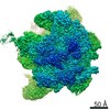
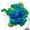
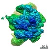
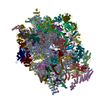
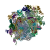
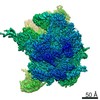
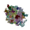
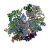
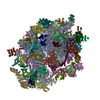
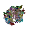
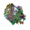
- PDB-6vmi: Structure of the human mitochondrial ribosome-EF-G1 complex (ClassIII) -
+ Open data

- Basic information
| Entry | Database: PDB / ID: 6vmi | ||||||
|---|---|---|---|---|---|---|---|
| Title | Structure of the human mitochondrial ribosome-EF-G1 complex (ClassIII) | ||||||
 Components Components |
| ||||||
 Keywords Keywords | RIBOSOME / mitochondrial elongation factor-G1 / 55S ribosome | ||||||
| Function / homology |  Function and homology information Function and homology informationmitochondrial transcription / mitochondrial translational termination / mitochondrial translational elongation / translation release factor activity, codon nonspecific / mitochondrial ribosome assembly / Mitochondrial translation elongation / Mitochondrial translation initiation / Mitochondrial ribosome-associated quality control / Mitochondrial translation termination / peptidyl-tRNA hydrolase ...mitochondrial transcription / mitochondrial translational termination / mitochondrial translational elongation / translation release factor activity, codon nonspecific / mitochondrial ribosome assembly / Mitochondrial translation elongation / Mitochondrial translation initiation / Mitochondrial ribosome-associated quality control / Mitochondrial translation termination / peptidyl-tRNA hydrolase / mitochondrial large ribosomal subunit / mitochondrial ribosome / mitochondrial small ribosomal subunit / peptidyl-tRNA hydrolase activity / mitochondrial translation / apoptotic mitochondrial changes / positive regulation of proteolysis / ribosomal small subunit binding / sperm head-tail coupling apparatus / anatomical structure morphogenesis / translation elongation factor activity / RNA processing / Mitochondrial protein degradation / rescue of stalled cytosolic ribosome / cellular response to leukemia inhibitory factor / apoptotic signaling pathway / fibrillar center / cell junction / regulation of translation / double-stranded RNA binding / large ribosomal subunit / endonuclease activity / small ribosomal subunit / small ribosomal subunit rRNA binding / large ribosomal subunit rRNA binding / nuclear membrane / Hydrolases; Acting on acid anhydrides; Acting on GTP to facilitate cellular and subcellular movement / tRNA binding / cell population proliferation / mitochondrial inner membrane / negative regulation of translation / rRNA binding / nuclear body / structural constituent of ribosome / ribosome / translation / mitochondrial matrix / ribonucleoprotein complex / protein domain specific binding / nucleotide binding / hydrolase activity / mRNA binding / GTPase activity / apoptotic process / positive regulation of DNA-templated transcription / GTP binding / nucleolus / mitochondrion / RNA binding / nucleoplasm / nucleus / plasma membrane / cytoplasm / cytosol Similarity search - Function | ||||||
| Biological species |  Homo sapiens (human) Homo sapiens (human) | ||||||
| Method | ELECTRON MICROSCOPY / single particle reconstruction / cryo EM / Resolution: 2.96 Å | ||||||
 Authors Authors | Sharma, M.R. / Koripella, R.K. / Agrawal, R.K. | ||||||
| Funding support |  United States, 1items United States, 1items
| ||||||
 Citation Citation | Journal: Nat Commun / Year: 2020 Title: Structures of the human mitochondrial ribosome bound to EF-G1 reveal distinct features of mitochondrial translation elongation. Authors: Ravi Kiran Koripella / Manjuli R Sharma / Kalpana Bhargava / Partha P Datta / Prem S Kaushal / Pooja Keshavan / Linda L Spremulli / Nilesh K Banavali / Rajendra K Agrawal /  Abstract: The mammalian mitochondrial ribosome (mitoribosome) and its associated translational factors have evolved to accommodate greater participation of proteins in mitochondrial translation. Here we ...The mammalian mitochondrial ribosome (mitoribosome) and its associated translational factors have evolved to accommodate greater participation of proteins in mitochondrial translation. Here we present the 2.68-3.96 Å cryo-EM structures of the human 55S mitoribosome in complex with the human mitochondrial elongation factor G1 (EF-G1) in three distinct conformational states, including an intermediate state and a post-translocational state. These structures reveal the role of several mitochondria-specific (mito-specific) mitoribosomal proteins (MRPs) and a mito-specific segment of EF-G1 in mitochondrial tRNA (tRNA) translocation. In particular, the mito-specific C-terminal extension in EF-G1 is directly involved in translocation of the acceptor arm of the A-site tRNA. In addition to the ratchet-like and independent head-swiveling motions exhibited by the small mitoribosomal subunit, we discover significant conformational changes in MRP mL45 at the nascent polypeptide-exit site within the large mitoribosomal subunit that could be critical for tethering of the elongating mitoribosome onto the inner-mitochondrial membrane. | ||||||
| History |
|
- Structure visualization
| Movie |
Movie viewer |
|---|---|
| Structure viewer | Molecule: MolmilJmol/JSmol |
- Downloads & links
-Download
| PDBx/mmCIF format | 6vmi.cif.gz | 3.9 MB | Display | PDBx/mmCIF format |
|---|---|---|---|---|
| PDB format | pdb6vmi.ent.gz | Display | PDB format | |
| PDBx/mmJSON format | 6vmi.json.gz | Tree view | PDBx/mmJSON format | |
| Others |  Other downloads Other downloads |
-Validation report
| Arichive directory | https://data.pdbj.org/pub/pdb/validation_reports/vm/6vmiftp://data.pdbj.org/pub/pdb/validation_reports/vm/6vmi | HTTPS FTP |
|---|
-Related structure data
| Related structure data | 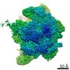 21242MC 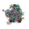 6vlzC C: citing same article ( M: map data used to model this data |
|---|---|
| Similar structure data | |
| EM raw data | EMPIAR-10481 (Title: Structures of the human mitochondrial ribosome bound to EF-G1 reveal distinct features of mitochondrial translation elongation Data size: 753.5 Data #1: Aligned single-frame particles of human mitochondrial 55S-EF-Gmt complex [picked particles - single frame - processed]) |


-Links
 PDBj
PDBj


- Assembly
| Deposited unit | 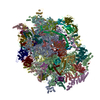
|
|---|---|
| 1 |
|
-Components
-RNA chain , 3 types, 3 molecules AAAB
| #1: RNA chain | Mass: 306112.125 Da / Num. of mol.: 1 / Source method: isolated from a natural source / Source: (natural) Homo sapiens (human) / References: GenBank: 1025814287 |
|---|---|
| #32: RNA chain | Mass: 500019.594 Da / Num. of mol.: 1 / Source method: isolated from a natural source / Source: (natural) Homo sapiens (human) / References: GenBank: 1563835895 |
| #33: RNA chain | Mass: 23266.883 Da / Num. of mol.: 1 / Source method: isolated from a natural source / Source: (natural) Homo sapiens (human) |
+28S ribosomal protein ... , 27 types, 27 molecules ABACAEAIAJAKAMANAOAPAQATAWAXAHALARASAUAVAYAZA1A0ADAFAG
-Protein , 10 types, 10 molecules A2A3A4joqPpuv
| #16: Protein | Mass: 13498.819 Da / Num. of mol.: 1 / Source method: isolated from a natural source / Source: (natural) Homo sapiens (human) / References: UniProt: Q96BP2 |
|---|---|
| #27: Protein | Mass: 22395.326 Da / Num. of mol.: 1 / Source method: isolated from a natural source / Source: (natural) Homo sapiens (human) / References: UniProt: Q9NWT8 |
| #28: Protein | Mass: 78648.547 Da / Num. of mol.: 1 / Source method: isolated from a natural source / Source: (natural) Homo sapiens (human) / References: UniProt: Q96EY7 |
| #58: Protein | Mass: 13696.684 Da / Num. of mol.: 1 / Source method: isolated from a natural source / Source: (natural) Homo sapiens (human) / References: UniProt: A8K7J6 |
| #60: Protein | Mass: 12292.333 Da / Num. of mol.: 1 / Source method: isolated from a natural source / Source: (natural) Homo sapiens (human) / References: UniProt: Q9BQC6 |
| #61: Protein | Mass: 25426.895 Da / Num. of mol.: 1 / Source method: isolated from a natural source / Source: (natural) Homo sapiens (human) / References: UniProt: Q8TAE8 |
| #66: Protein | Mass: 20465.348 Da / Num. of mol.: 1 / Source method: isolated from a natural source / Source: (natural) Homo sapiens (human) / References: UniProt: A8K9D2 |
| #81: Protein | Mass: 23674.203 Da / Num. of mol.: 1 / Source method: isolated from a natural source / Source: (natural) Homo sapiens (human) / References: UniProt: Q14197, peptidyl-tRNA hydrolase |
| #85: Protein | Mass: 5549.833 Da / Num. of mol.: 1 / Source method: isolated from a natural source / Source: (natural) Homo sapiens (human) |
| #86: Protein | Mass: 83578.461 Da / Num. of mol.: 1 / Source method: isolated from a natural source / Source: (natural) Homo sapiens (human) / References: UniProt: Q96RP9 |
+39S ribosomal protein ... , 46 types, 48 molecules DFHKLMORSTWXYZ012348begimrJINU...
-DNA chain , 2 types, 3 molecules A5A6A7
| #87: DNA chain | Mass: 3183.098 Da / Num. of mol.: 1 / Source method: isolated from a natural source / Source: (natural) Homo sapiens (human) |
|---|---|
| #88: DNA chain | Mass: 20742.291 Da / Num. of mol.: 2 / Source method: isolated from a natural source / Source: (natural) Homo sapiens (human) |
-Non-polymers , 4 types, 139 molecules 


| #89: Chemical | ChemComp-MG /  Mass: 24.305 Da / Num. of mol.: 130 / Source method: obtained synthetically / Formula: Mg Mass: 24.305 Da / Num. of mol.: 130 / Source method: obtained synthetically / Formula: Mg#90: Chemical | ChemComp-ZN /  Mass: 65.409 Da / Num. of mol.: 7 / Source method: obtained synthetically / Formula: Zn Mass: 65.409 Da / Num. of mol.: 7 / Source method: obtained synthetically / Formula: Zn#91: Chemical | ChemComp-GDP / |  Type: RNA linking / Mass: 443.201 Da / Num. of mol.: 1 / Source method: obtained synthetically / Formula: C10H15N5O11P2 / Comment: GDP, energy-carrying molecule*YM Type: RNA linking / Mass: 443.201 Da / Num. of mol.: 1 / Source method: obtained synthetically / Formula: C10H15N5O11P2 / Comment: GDP, energy-carrying molecule*YM#92: Chemical | ChemComp-GCP / |  Mass: 521.208 Da / Num. of mol.: 1 / Source method: obtained synthetically / Formula: C11H18N5O13P3 / Comment: GMP-PCP, energy-carrying molecule analogue*YM Mass: 521.208 Da / Num. of mol.: 1 / Source method: obtained synthetically / Formula: C11H18N5O13P3 / Comment: GMP-PCP, energy-carrying molecule analogue*YM |
|---|
-Details
| Has ligand of interest | N |
|---|---|
| Has protein modification | Y |
-Experimental details
-Experiment
| Experiment | Method: ELECTRON MICROSCOPY |
|---|---|
| EM experiment | Aggregation state: PARTICLE / 3D reconstruction method: single particle reconstruction |
- Sample preparation
| Component | Name: Human mitochondrial ribosome-EF-G1 complex / Type: COMPLEX / Entity ID: #1-#88 / Source: MULTIPLE SOURCES |
|---|---|
| Source (natural) | Organism: Homo sapiens (human) |
| Buffer solution | pH: 7.5 |
| Specimen | Embedding applied: NO / Shadowing applied: NO / Staining applied: NO / Vitrification applied: YES |
| Vitrification | Cryogen name: ETHANE |
- Electron microscopy imaging
| Experimental equipment |  Model: Titan Krios / Image courtesy: FEI Company |
|---|---|
| Microscopy | Model: FEI TITAN KRIOS |
| Electron gun | Electron source:  FIELD EMISSION GUN / Accelerating voltage: 300 kV / Illumination mode: FLOOD BEAM FIELD EMISSION GUN / Accelerating voltage: 300 kV / Illumination mode: FLOOD BEAM |
| Electron lens | Mode: BRIGHT FIELD |
| Image recording | Electron dose: 69.2 e/Å2 / Detector mode: COUNTING / Film or detector model: GATAN K2 SUMMIT (4k x 4k) |
- Processing
| CTF correction | Type: PHASE FLIPPING AND AMPLITUDE CORRECTION |
|---|---|
| 3D reconstruction | Resolution: 2.96 Å / Resolution method: FSC 0.143 CUT-OFF / Num. of particles: 150347 / Symmetry type: POINT |
