 Movie
Movie Controller

[English] 日本語
 Yorodumi
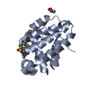
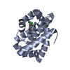
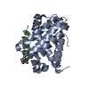
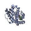
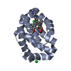
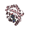
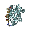
- PDB-6vbx: Crystal structure of Mcl-1 in complex with 138E12 peptide, Lys-co... -
+ Open data

- Basic information
| Entry | Database: PDB / ID: 6vbx | ||||||
|---|---|---|---|---|---|---|---|
| Title | Crystal structure of Mcl-1 in complex with 138E12 peptide, Lys-covalent antagonist | ||||||
 Components Components |
| ||||||
 Keywords Keywords | APOPTOSIS / MCL-1 / 138E12 peptide / Lys-covalent antagonists | ||||||
| Function / homology |  Function and homology information Function and homology informationBIM-BCL-xl complex / BIM-BCL-2 complex / regulation of developmental pigmentation / RUNX3 regulates BCL2L11 (BIM) transcription / positive regulation of mitochondrial membrane permeability involved in apoptotic process / positive regulation of endoplasmic reticulum stress-induced intrinsic apoptotic signaling pathway / positive regulation of fibroblast apoptotic process / Activation of BIM and translocation to mitochondria / apoptotic process involved in embryonic digit morphogenesis / developmental pigmentation ...BIM-BCL-xl complex / BIM-BCL-2 complex / regulation of developmental pigmentation / RUNX3 regulates BCL2L11 (BIM) transcription / positive regulation of mitochondrial membrane permeability involved in apoptotic process / positive regulation of endoplasmic reticulum stress-induced intrinsic apoptotic signaling pathway / positive regulation of fibroblast apoptotic process / Activation of BIM and translocation to mitochondria / apoptotic process involved in embryonic digit morphogenesis / developmental pigmentation / ear development / positive regulation of oxidative stress-induced neuron intrinsic apoptotic signaling pathway / BH3-only proteins associate with and inactivate anti-apoptotic BCL-2 members / cell fate determination / positive regulation of T cell apoptotic process / tube formation / regulation of organ growth / mammary gland development / cellular homeostasis / meiosis I / mitochondrial fusion / myeloid cell homeostasis / Bcl-2 family protein complex / cellular response to glucocorticoid stimulus / NRAGE signals death through JNK / thymocyte apoptotic process / Deregulated CDK5 triggers multiple neurodegenerative pathways in Alzheimer's disease models / FOXO-mediated transcription of cell death genes / positive regulation of IRE1-mediated unfolded protein response / odontogenesis of dentin-containing tooth / B cell homeostasis / T cell homeostasis / positive regulation of release of cytochrome c from mitochondria / negative regulation of anoikis / BH3 domain binding / transmembrane protein transporter activity / negative regulation of extrinsic apoptotic signaling pathway in absence of ligand / spleen development / extrinsic apoptotic signaling pathway in absence of ligand / positive regulation of intrinsic apoptotic signaling pathway / positive regulation of cell cycle / endomembrane system / FLT3 Signaling / response to cytokine / release of cytochrome c from mitochondria / response to endoplasmic reticulum stress / thymus development / cell-matrix adhesion / negative regulation of autophagy / post-embryonic development / kidney development / positive regulation of protein-containing complex assembly / male gonad development / intrinsic apoptotic signaling pathway in response to DNA damage / Signaling by BRAF and RAF1 fusions / Signaling by ALK fusions and activated point mutants / positive regulation of neuron apoptotic process / channel activity / Interleukin-4 and Interleukin-13 signaling / spermatogenesis / regulation of apoptotic process / microtubule binding / in utero embryonic development / mitochondrial outer membrane / positive regulation of apoptotic process / protein heterodimerization activity / apoptotic process / DNA damage response / protein kinase binding / negative regulation of apoptotic process / mitochondrion / nucleoplasm / membrane / nucleus / cytoplasm / cytosol Similarity search - Function | ||||||
| Biological species |  Homo sapiens (human) Homo sapiens (human)synthetic construct (others) | ||||||
| Method |  X-RAY DIFFRACTION / SYNCHROTRON / MOLECULAR REPLACEMENT / Resolution: 1.95 Å X-RAY DIFFRACTION / SYNCHROTRON / MOLECULAR REPLACEMENT / Resolution: 1.95 Å | ||||||
 Authors Authors | Pellecchia, M. / Perry, J.J. / Kenjic, N. / Assar, Z. | ||||||
| Funding support |  United States, 1items United States, 1items
| ||||||
 Citation Citation | Journal: J.Med.Chem. / Year: 2021 Title: Design, Synthesis, and Structural Characterization of Lysine Covalent BH3 Peptides Targeting Mcl-1. Authors: Gambini, L. / Udompholkul, P. / Baggio, C. / Muralidharan, A. / Kenjic, N. / Assar, Z. / Perry, J.J.P. / Pellecchia, M. #1: Journal: Meth. Enzymol. / Year: 1997 Title: Processing of X-ray diffraction data collected in oscillation mode. Authors: Otwinowski, Z. / Minor, W. #2: Journal: J Appl Crystallogr / Year: 2007 Title: Phaser crystallographic software. Authors: McCoy, A.J. / Grosse-Kunstleve, R.W. / Adams, P.D. / Winn, M.D. / Storoni, L.C. / Read, R.J. #3: Journal: Acta Crystallogr. D Biol. Crystallogr. / Year: 2004 Title: REFMAC5 dictionary: organization of prior chemical knowledge and guidelines for its use. Authors: Vagin, A.A. / Steiner, R.A. / Lebedev, A.A. / Potterton, L. / McNicholas, S. / Long, F. / Murshudov, G.N. #4: Journal: Acta Crystallogr D Biol Crystallogr / Year: 2004 Title: Coot: model-building tools for molecular graphics. Authors: Paul Emsley / Kevin Cowtan /  Abstract: CCP4mg is a project that aims to provide a general-purpose tool for structural biologists, providing tools for X-ray structure solution, structure comparison and analysis, and publication-quality ...CCP4mg is a project that aims to provide a general-purpose tool for structural biologists, providing tools for X-ray structure solution, structure comparison and analysis, and publication-quality graphics. The map-fitting tools are available as a stand-alone package, distributed as 'Coot'. | ||||||
| History |
|
- Structure visualization
| Structure viewer | Molecule: MolmilJmol/JSmol |
|---|
- Downloads & links
-Download
| PDBx/mmCIF format | 6vbx.cif.gz | 56.3 KB | Display | PDBx/mmCIF format |
|---|---|---|---|---|
| PDB format | pdb6vbx.ent.gz | 31.4 KB | Display | PDB format |
| PDBx/mmJSON format | 6vbx.json.gz | Tree view | PDBx/mmJSON format | |
| Others |  Other downloads Other downloads |
-Validation report
| Arichive directory | https://data.pdbj.org/pub/pdb/validation_reports/vb/6vbxftp://data.pdbj.org/pub/pdb/validation_reports/vb/6vbx | HTTPS FTP |
|---|
-Related structure data
| Related structure data |  2nl9S S: Starting model for refinement |
|---|---|
| Similar structure data |


-Links
 PDBj
PDBj


- Assembly
| Deposited unit | 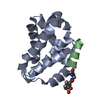
| ||||||||||
|---|---|---|---|---|---|---|---|---|---|---|---|
| 1 |
| ||||||||||
| 2 | 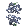
| ||||||||||
| Unit cell |
|
-Components
| #1: Protein | Mass: 17850.330 Da / Num. of mol.: 1 Source method: isolated from a genetically manipulated source Source: (gene. exp.) Homo sapiens (human) / Gene: MCL1, BCL2L3 / Production host:  |
|---|---|
| #2: Protein/peptide | Mass: 1805.020 Da / Num. of mol.: 1 / Source method: obtained synthetically Details: This is a chemically synthesized peptide with unnatural ligand (see ligand section) on the N-terminal end. Ligands is covalently attached to Lys234 of chain A. Source: (synth.) synthetic construct (others) / References: UniProt: O43521*PLUS |
| #3: Water | ChemComp-HOH /  Mass: 18.015 Da / Num. of mol.: 44 / Source method: isolated from a natural source / Formula: H2O Mass: 18.015 Da / Num. of mol.: 44 / Source method: isolated from a natural source / Formula: H2O |
| Has ligand of interest | Y |
-Experimental details
-Experiment
| Experiment | Method: X-RAY DIFFRACTION / Number of used crystals: 1 |
|---|
- Sample preparation
| Crystal | Density Matthews: 2.18 Å3/Da / Density % sol: 43.55 % |
|---|---|
| Crystal grow | Temperature: 298 K / Method: vapor diffusion, sitting drop Details: 0.5M Potassium Thiocyanate, 0.1M Sodium Acetate:HCL pH4.6 |
-Data collection
| Diffraction | Mean temperature: 100 K / Serial crystal experiment: N |
|---|---|
| Diffraction source | Source: SYNCHROTRON / Site: APS / Beamline: 21-ID-D / Wavelength: 1.127 Å |
| Detector | Type: DECTRIS EIGER X 9M / Detector: PIXEL / Date: Oct 31, 2019 |
| Radiation | Protocol: SINGLE WAVELENGTH / Monochromatic (M) / Laue (L): M / Scattering type: x-ray |
| Radiation wavelength | Wavelength: 1.127 Å / Relative weight: 1 |
| Reflection | Resolution: 1.95→34.001 Å / Num. obs: 12721 / % possible obs: 99.75 % / Redundancy: 7.9 % / Biso Wilson estimate: 32.63 Å2 / Rmerge(I) obs: 0.073 / Net I/σ(I): 22 |
| Reflection shell | Resolution: 1.955→2.025 Å / Redundancy: 8.4 % / Rmerge(I) obs: 0.276 / Mean I/σ(I) obs: 2.3 / Num. unique obs: 1235 / % possible all: 100 |
- Processing
| Software |
| ||||||||||||||||||||||||||||
|---|---|---|---|---|---|---|---|---|---|---|---|---|---|---|---|---|---|---|---|---|---|---|---|---|---|---|---|---|---|
| Refinement | Method to determine structure: MOLECULAR REPLACEMENT Starting model: 2NL9 Resolution: 1.95→34 Å / SU ML: 0.2912 / Cross valid method: FREE R-VALUE / σ(F): 1.34 / Phase error: 34.0423 Stereochemistry target values: GeoStd + Monomer Library + CDL v1.2
| ||||||||||||||||||||||||||||
| Solvent computation | Shrinkage radii: 0.9 Å / VDW probe radii: 1.11 Å / Solvent model: FLAT BULK SOLVENT MODEL | ||||||||||||||||||||||||||||
| Displacement parameters | Biso mean: 35.58 Å2 | ||||||||||||||||||||||||||||
| Refinement step | Cycle: LAST / Resolution: 1.95→34 Å
| ||||||||||||||||||||||||||||
| Refine LS restraints |
| ||||||||||||||||||||||||||||
| LS refinement shell |
|
