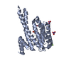
 Movie
Movie Controller

[English] 日本語
 Yorodumi
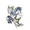
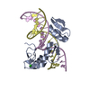
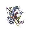
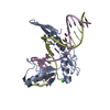
- PDB-6v8u: Kaiso (ZBTB33) zinc finger DNA binding domain in complex with a m... -
+ Open data

- Basic information
| Entry | Database: PDB / ID: 6v8u | ||||||
|---|---|---|---|---|---|---|---|
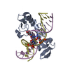
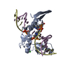
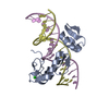
| Title | Kaiso (ZBTB33) zinc finger DNA binding domain in complex with a modified Kaiso binding sequence (KBS) | ||||||
 Components Components |
| ||||||
 Keywords Keywords | DNA BINDING PROTEIN / DNA methylation / Zinc finger / Transcriptional regulator | ||||||
| Function / homology |  Function and homology information Function and homology informationmethyl-CpG binding / regulation of immune system process / regulation of cytokine production / Negative Regulation of CDH1 Gene Transcription / DNA-binding transcription repressor activity, RNA polymerase II-specific / Wnt signaling pathway / sequence-specific double-stranded DNA binding / sequence-specific DNA binding / DNA-binding transcription factor activity, RNA polymerase II-specific / intracellular signal transduction ...methyl-CpG binding / regulation of immune system process / regulation of cytokine production / Negative Regulation of CDH1 Gene Transcription / DNA-binding transcription repressor activity, RNA polymerase II-specific / Wnt signaling pathway / sequence-specific double-stranded DNA binding / sequence-specific DNA binding / DNA-binding transcription factor activity, RNA polymerase II-specific / intracellular signal transduction / RNA polymerase II cis-regulatory region sequence-specific DNA binding / negative regulation of DNA-templated transcription / chromatin / negative regulation of transcription by RNA polymerase II / DNA-templated transcription / zinc ion binding / nucleoplasm / nucleus / plasma membrane / cytosol Similarity search - Function | ||||||
| Biological species |  Homo sapiens (human) Homo sapiens (human) | ||||||
| Method |  X-RAY DIFFRACTION / SYNCHROTRON / MOLECULAR REPLACEMENT / Resolution: 2.103 Å X-RAY DIFFRACTION / SYNCHROTRON / MOLECULAR REPLACEMENT / Resolution: 2.103 Å | ||||||
 Authors Authors | Nikolova, E.N. / Stanfield, R.L. / Dyson, H.J. / Wright, P.E. | ||||||
| Funding support |  United States, 1items United States, 1items
| ||||||
 Citation Citation | Journal: Biochemistry / Year: 2020 Title: A Conformational Switch in the Zinc Finger Protein Kaiso Mediates Differential Readout of Specific and Methylated DNA Sequences. Authors: Nikolova, E.N. / Stanfield, R.L. / Dyson, H.J. / Wright, P.E. | ||||||
| History |
|
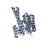
- Structure visualization
| Structure viewer | Molecule: MolmilJmol/JSmol |
|---|
- Downloads & links
-Download
| PDBx/mmCIF format | 6v8u.cif.gz | 116.7 KB | Display | PDBx/mmCIF format |
|---|---|---|---|---|
| PDB format | pdb6v8u.ent.gz | 84.9 KB | Display | PDB format |
| PDBx/mmJSON format | 6v8u.json.gz | Tree view | PDBx/mmJSON format | |
| Others |  Other downloads Other downloads |
-Validation report
| Arichive directory | https://data.pdbj.org/pub/pdb/validation_reports/v8/6v8uftp://data.pdbj.org/pub/pdb/validation_reports/v8/6v8u | HTTPS FTP |
|---|
-Related structure data
| Related structure data |  6df5C  6df8C  6df9C  6dfaC  6dfbC  6dfcC 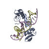 4f6mS C: citing same article ( S: Starting model for refinement |
|---|---|
| Similar structure data |

-Links
 PDBj
PDBj


- Assembly
| Deposited unit | 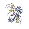
| ||||||||||
|---|---|---|---|---|---|---|---|---|---|---|---|
| 1 |
| ||||||||||
| Unit cell |
| ||||||||||
| Components on special symmetry positions |
|
-Components
-Protein , 1 types, 1 molecules A
| #1: Protein | Mass: 16217.771 Da / Num. of mol.: 1 Source method: isolated from a genetically manipulated source Source: (gene. exp.) Homo sapiens (human) / Gene: ZBTB33, KAISO, ZNF348 / Plasmid: PET21D / Production host:  |
|---|
-DNA chain , 2 types, 2 molecules DE
| #2: DNA chain | Mass: 5491.566 Da / Num. of mol.: 1 / Source method: obtained synthetically / Source: (synth.) Homo sapiens (human) |
|---|---|
| #3: DNA chain | Mass: 5540.604 Da / Num. of mol.: 1 / Source method: obtained synthetically / Source: (synth.) Homo sapiens (human) |
-Non-polymers , 4 types, 112 molecules 


| #4: Chemical |  Mass: 65.409 Da / Num. of mol.: 3 / Source method: obtained synthetically / Formula: Zn Mass: 65.409 Da / Num. of mol.: 3 / Source method: obtained synthetically / Formula: Zn#5: Chemical | ChemComp-CL / |  Mass: 35.453 Da / Num. of mol.: 1 / Source method: obtained synthetically / Formula: Cl Mass: 35.453 Da / Num. of mol.: 1 / Source method: obtained synthetically / Formula: Cl#6: Chemical | ChemComp-NA / |  Mass: 22.990 Da / Num. of mol.: 1 / Source method: isolated from a natural source / Formula: Na Mass: 22.990 Da / Num. of mol.: 1 / Source method: isolated from a natural source / Formula: Na#7: Water | ChemComp-HOH / | Mass: 18.015 Da / Num. of mol.: 107 / Source method: isolated from a natural source / Formula: H2O |
|---|
-Details
| Has ligand of interest | N |
|---|
-Experimental details
-Experiment
| Experiment | Method: X-RAY DIFFRACTION / Number of used crystals: 1 |
|---|
- Sample preparation
| Crystal | Density Matthews: 3.62 Å3/Da / Density % sol: 65.98 % |
|---|---|
| Crystal grow | Temperature: 295 K / Method: vapor diffusion, sitting drop / pH: 5 Details: 0.02 M Calcium Chloride, 0.1 M Sodium Acetate, 30 % 2-methyl-2,4-pentanediol |
-Data collection
| Diffraction | Mean temperature: 100 K / Serial crystal experiment: N |
|---|---|
| Diffraction source | Source: SYNCHROTRON / Site: SSRL / Beamline: BL12-2 / Wavelength: 0.9795 Å |
| Detector | Type: DECTRIS PILATUS 6M / Detector: PIXEL / Date: Mar 18, 2014 |
| Radiation | Protocol: SINGLE WAVELENGTH / Monochromatic (M) / Laue (L): M / Scattering type: x-ray |
| Radiation wavelength | Wavelength: 0.9795 Å / Relative weight: 1 |
| Reflection | Resolution: 2.103→42.92 Å / Num. obs: 23073 / % possible obs: 97.56 % / Redundancy: 11.1 % / Biso Wilson estimate: 38.8250910295 Å2 / CC1/2: 0.996 / CC star: 0.999 / Rmerge(I) obs: 0.1071 / Rrim(I) all: 0.1123 / Net I/σ(I): 17.6 |
| Reflection shell | Resolution: 2.103→2.178 Å / Redundancy: 6.6 % / Rmerge(I) obs: 0.7772 / Mean I/σ(I) obs: 2.83 / Num. unique obs: 1986 / CC1/2: 0.86 / CC star: 0.961 / % possible all: 86.23 |
- Processing
| Software |
| ||||||||||||||||||||||||||||||||||||||||||||||||||||||||||||||||||||||||||||||||||||||||||||||||||||||||||||||||||||||||||||||||||||||||||||||||||||||||||||||||||||||||||||||||||||||||||||||||||||||||
|---|---|---|---|---|---|---|---|---|---|---|---|---|---|---|---|---|---|---|---|---|---|---|---|---|---|---|---|---|---|---|---|---|---|---|---|---|---|---|---|---|---|---|---|---|---|---|---|---|---|---|---|---|---|---|---|---|---|---|---|---|---|---|---|---|---|---|---|---|---|---|---|---|---|---|---|---|---|---|---|---|---|---|---|---|---|---|---|---|---|---|---|---|---|---|---|---|---|---|---|---|---|---|---|---|---|---|---|---|---|---|---|---|---|---|---|---|---|---|---|---|---|---|---|---|---|---|---|---|---|---|---|---|---|---|---|---|---|---|---|---|---|---|---|---|---|---|---|---|---|---|---|---|---|---|---|---|---|---|---|---|---|---|---|---|---|---|---|---|---|---|---|---|---|---|---|---|---|---|---|---|---|---|---|---|---|---|---|---|---|---|---|---|---|---|---|---|---|---|---|---|---|
| Refinement | Method to determine structure: MOLECULAR REPLACEMENT Starting model: 4F6M Resolution: 2.103→42.919 Å / SU ML: 0.21 / Cross valid method: THROUGHOUT / σ(F): 1.33 / Phase error: 22.07 / Stereochemistry target values: ML
| ||||||||||||||||||||||||||||||||||||||||||||||||||||||||||||||||||||||||||||||||||||||||||||||||||||||||||||||||||||||||||||||||||||||||||||||||||||||||||||||||||||||||||||||||||||||||||||||||||||||||
| Solvent computation | Shrinkage radii: 0.9 Å / VDW probe radii: 1.11 Å / Solvent model: FLAT BULK SOLVENT MODEL | ||||||||||||||||||||||||||||||||||||||||||||||||||||||||||||||||||||||||||||||||||||||||||||||||||||||||||||||||||||||||||||||||||||||||||||||||||||||||||||||||||||||||||||||||||||||||||||||||||||||||
| Displacement parameters | Biso max: 137.62 Å2 / Biso mean: 56.4997 Å2 / Biso min: 22.26 Å2 | ||||||||||||||||||||||||||||||||||||||||||||||||||||||||||||||||||||||||||||||||||||||||||||||||||||||||||||||||||||||||||||||||||||||||||||||||||||||||||||||||||||||||||||||||||||||||||||||||||||||||
| Refinement step | Cycle: final / Resolution: 2.103→42.919 Å
| ||||||||||||||||||||||||||||||||||||||||||||||||||||||||||||||||||||||||||||||||||||||||||||||||||||||||||||||||||||||||||||||||||||||||||||||||||||||||||||||||||||||||||||||||||||||||||||||||||||||||
| LS refinement shell | Refine-ID: X-RAY DIFFRACTION / Rfactor Rfree error: 0
| ||||||||||||||||||||||||||||||||||||||||||||||||||||||||||||||||||||||||||||||||||||||||||||||||||||||||||||||||||||||||||||||||||||||||||||||||||||||||||||||||||||||||||||||||||||||||||||||||||||||||
| Refinement TLS params. | Method: refined / Refine-ID: X-RAY DIFFRACTION
| ||||||||||||||||||||||||||||||||||||||||||||||||||||||||||||||||||||||||||||||||||||||||||||||||||||||||||||||||||||||||||||||||||||||||||||||||||||||||||||||||||||||||||||||||||||||||||||||||||||||||
| Refinement TLS group |
|
