 Movie
Movie Controller

[English] 日本語
 Yorodumi
- PDB-6v5c: Human Drosha and DGCR8 in complex with Primary MicroRNA (MP/RNA c... -
+ Open data

- Basic information
| Entry | Database: PDB / ID: 6v5c | |||||||||||||||
|---|---|---|---|---|---|---|---|---|---|---|---|---|---|---|---|---|
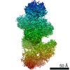
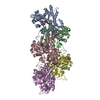
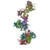
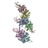
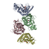
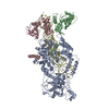
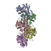
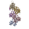
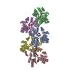
| Title | Human Drosha and DGCR8 in complex with Primary MicroRNA (MP/RNA complex) - partially docked state | |||||||||||||||
 Components Components |
| |||||||||||||||
 Keywords Keywords | RNA BINDING PROTEIN/RNA / Microprocessor / Drosha / DGCR8 / Primary MicroRNA / RNA BINDING PROTEIN / RNA BINDING PROTEIN-RNA complex | |||||||||||||||
| Function / homology |  Function and homology information Function and homology informationpositive regulation of pre-miRNA processing / microprocessor complex / regulation of miRNA metabolic process / DEAD/H-box RNA helicase binding / protein-RNA adaptor activity / primary miRNA binding / ribonuclease III / regulation of regulatory T cell differentiation / Transcriptional Regulation by MECP2 / regulation of stem cell proliferation ...positive regulation of pre-miRNA processing / microprocessor complex / regulation of miRNA metabolic process / DEAD/H-box RNA helicase binding / protein-RNA adaptor activity / primary miRNA binding / ribonuclease III / regulation of regulatory T cell differentiation / Transcriptional Regulation by MECP2 / regulation of stem cell proliferation / primary miRNA processing / miRNA metabolic process / ribonuclease III activity / pre-miRNA processing / MicroRNA (miRNA) biogenesis / SMAD binding / R-SMAD binding / lipopolysaccharide binding / rRNA processing / double-stranded RNA binding / site of double-strand break / regulation of inflammatory response / defense response to Gram-negative bacterium / protein-macromolecule adaptor activity / postsynaptic density / defense response to Gram-positive bacterium / nuclear body / heme binding / DNA damage response / positive regulation of gene expression / nucleolus / glutamatergic synapse / protein homodimerization activity / RNA binding / nucleoplasm / metal ion binding / identical protein binding / nucleus / cytoplasm / cytosol Similarity search - Function | |||||||||||||||
| Biological species |  Homo sapiens (human) Homo sapiens (human) | |||||||||||||||
| Method | ELECTRON MICROSCOPY / single particle reconstruction / cryo EM / Resolution: 4.4 Å | |||||||||||||||
 Authors Authors | Partin, A. / Zhang, K. / Jeong, B. / Herrell, E. / Li, S. / Chiu, W. / Nam, Y. | |||||||||||||||
| Funding support |  United States, 4items United States, 4items
| |||||||||||||||
 Citation Citation | Journal: Mol Cell / Year: 2020 Title: Cryo-EM Structures of Human Drosha and DGCR8 in Complex with Primary MicroRNA. Authors: Alexander C Partin / Kaiming Zhang / Byung-Cheon Jeong / Emily Herrell / Shanshan Li / Wah Chiu / Yunsun Nam / Abstract: Metazoan microRNAs require specific maturation steps initiated by Microprocessor, comprising Drosha and DGCR8. Lack of structural information for the assembled complex has hindered an understanding ...Metazoan microRNAs require specific maturation steps initiated by Microprocessor, comprising Drosha and DGCR8. Lack of structural information for the assembled complex has hindered an understanding of how Microprocessor recognizes primary microRNA transcripts (pri-miRNAs). Here we present a cryoelectron microscopy structure of human Microprocessor with a pri-miRNA docked in the active site, poised for cleavage. The basal junction is recognized by a four-way intramolecular junction in Drosha, triggered by the Belt and Wedge regions that clamp over the ssRNA. The belt is important for efficiency and accuracy of pri-miRNA processing. Two dsRBDs form a molecular ruler to measure the stem length between the two dsRNA-ssRNA junctions. The specific organization of the dsRBDs near the apical junction is independent of Drosha core domains, as observed in a second structure in the partially docked state. Collectively, we derive a molecular model to explain how Microprocessor recognizes a pri-miRNA and accurately identifies the cleavage site. | |||||||||||||||
| History |
|
- Structure visualization
| Movie |
Movie viewer |
|---|---|
| Structure viewer | Molecule: MolmilJmol/JSmol |
- Downloads & links
-Download
| PDBx/mmCIF format | 6v5c.cif.gz | 302 KB | Display | PDBx/mmCIF format |
|---|---|---|---|---|
| PDB format | pdb6v5c.ent.gz | 227.1 KB | Display | PDB format |
| PDBx/mmJSON format | 6v5c.json.gz | Tree view | PDBx/mmJSON format | |
| Others |  Other downloads Other downloads |
-Validation report
| Arichive directory | https://data.pdbj.org/pub/pdb/validation_reports/v5/6v5cftp://data.pdbj.org/pub/pdb/validation_reports/v5/6v5c | HTTPS FTP |
|---|
-Related structure data
| Related structure data | 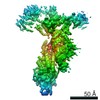 21052MC  6v5bC C: citing same article ( M: map data used to model this data |
|---|---|
| Similar structure data |


-Links
 PDBj
PDBj


- Assembly
| Deposited unit | 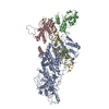
|
|---|---|
| 1 |
|
-Components
| #1: Protein | Mass: 118377.742 Da / Num. of mol.: 1 / Mutation: E1045Q, E1222Q Source method: isolated from a genetically manipulated source Source: (gene. exp.) Homo sapiens (human) / Gene: DROSHA, RN3, RNASE3L, RNASEN / Production host:  Baculovirus expression vector pFastBac1-HM / References: UniProt: Q9NRR4, ribonuclease III Baculovirus expression vector pFastBac1-HM / References: UniProt: Q9NRR4, ribonuclease III | ||
|---|---|---|---|
| #2: Protein | Mass: 60550.602 Da / Num. of mol.: 2 Source method: isolated from a genetically manipulated source Source: (gene. exp.) Homo sapiens (human) / Gene: DGCR8, C22orf12, DGCRK6, LP4941Production host:  References: UniProt: Q8WYQ5 #3: RNA chain | | Mass: 33646.934 Da / Num. of mol.: 1 Source method: isolated from a genetically manipulated source Source: (gene. exp.) Homo sapiens (human) / Production host: Transcription vector pUC18-pES2 (others) / References: GenBank: 21206011 |
-Experimental details
-Experiment
| Experiment | Method: ELECTRON MICROSCOPY |
|---|---|
| EM experiment | Aggregation state: PARTICLE / 3D reconstruction method: single particle reconstruction |
- Sample preparation
| Component | Name: Human Drosha and DGCR8 in complex with Primary MicroRNA (MP/RNA complex) - partially docked state Type: COMPLEX / Entity ID: all / Source: MULTIPLE SOURCES | ||||||||||||
|---|---|---|---|---|---|---|---|---|---|---|---|---|---|
| Molecular weight |
| ||||||||||||
| Source (natural) | Organism: Homo sapiens (human) | ||||||||||||
| Buffer solution | pH: 7.1 | ||||||||||||
| Specimen | Conc.: 3.7 mg/ml / Embedding applied: NO / Shadowing applied: NO / Staining applied: NO / Vitrification applied: YES Details: Human Drosha and DGCR8 in complex with Primary MicroRNA (MP/RNA complex) - partially docked state | ||||||||||||
| Vitrification | Instrument: FEI VITROBOT MARK IV / Cryogen name: ETHANE / Humidity: 100 % |
- Electron microscopy imaging
| Experimental equipment |  Model: Titan Krios / Image courtesy: FEI Company |
|---|---|
| Microscopy | Model: FEI TITAN KRIOS |
| Electron gun | Electron source:  FIELD EMISSION GUN / Accelerating voltage: 300 kV / Illumination mode: FLOOD BEAM FIELD EMISSION GUN / Accelerating voltage: 300 kV / Illumination mode: FLOOD BEAM |
| Electron lens | Mode: BRIGHT FIELD / Nominal magnification: 130000 X / Nominal defocus max: 3600 nm / Nominal defocus min: 1500 nm / Cs: 2.7 mm / C2 aperture diameter: 70 µm / Alignment procedure: COMA FREE |
| Specimen holder | Cryogen: NITROGEN / Specimen holder model: FEI TITAN KRIOS AUTOGRID HOLDER |
| Image recording | Average exposure time: 6 sec. / Electron dose: 46.8 e/Å2 / Detector mode: COUNTING / Film or detector model: GATAN K2 SUMMIT (4k x 4k) / Num. of grids imaged: 4 / Num. of real images: 6070 |
| EM imaging optics | Energyfilter name: GIF Quantum LS / Energyfilter slit width: 20 eV |
| Image scans | Movie frames/image: 30 / Used frames/image: 1-30 |
- Processing
| Software | Name: PHENIX / Version: 1.14_3260: / Classification: refinement | ||||||||||||||||||||||||||||||||||||
|---|---|---|---|---|---|---|---|---|---|---|---|---|---|---|---|---|---|---|---|---|---|---|---|---|---|---|---|---|---|---|---|---|---|---|---|---|---|
| EM software |
| ||||||||||||||||||||||||||||||||||||
| CTF correction | Type: PHASE FLIPPING AND AMPLITUDE CORRECTION | ||||||||||||||||||||||||||||||||||||
| Particle selection | Num. of particles selected: 1063710 | ||||||||||||||||||||||||||||||||||||
| Symmetry | Point symmetry: C1 (asymmetric) | ||||||||||||||||||||||||||||||||||||
| 3D reconstruction | Resolution: 4.4 Å / Resolution method: FSC 0.143 CUT-OFF / Num. of particles: 381468 / Symmetry type: POINT | ||||||||||||||||||||||||||||||||||||
| Atomic model building | Protocol: BACKBONE TRACE / Space: REAL | ||||||||||||||||||||||||||||||||||||
| Refine LS restraints |
|
