 Movie
Movie Controller

[English] 日本語
 Yorodumi
- PDB-6v1x: Cryo-EM Structure of the Hyperpolarization-Activated Potassium Ch... -
+ Open data

- Basic information
| Entry | Database: PDB / ID: 6v1x | |||||||||
|---|---|---|---|---|---|---|---|---|---|---|
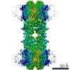
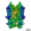
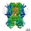
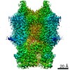
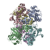
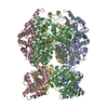
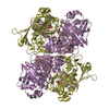
| Title | Cryo-EM Structure of the Hyperpolarization-Activated Potassium Channel KAT1: Tetramer | |||||||||
 Components Components | Potassium channel KAT1 | |||||||||
 Keywords Keywords | TRANSPORT PROTEIN / membrane protein / voltage-gated ion channel / potassium channel | |||||||||
| Function / homology |  Function and homology information Function and homology informationinward rectifier potassium channel activity / monoatomic ion channel complex / identical protein binding / plasma membrane Similarity search - Function | |||||||||
| Biological species |  | |||||||||
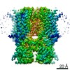
| Method | ELECTRON MICROSCOPY / single particle reconstruction / cryo EM / Resolution: 3.5 Å | |||||||||
 Authors Authors | Clark, M.D. / Contreras, G.F. / Shen, R. / Perozo, E. | |||||||||
| Funding support |  United States, 2items United States, 2items
| |||||||||
 Citation Citation | Journal: Nature / Year: 2020 Title: Electromechanical coupling in the hyperpolarization-activated K channel KAT1. Authors: Michael David Clark / Gustavo F Contreras / Rong Shen / Eduardo Perozo / Abstract: Voltage-gated potassium (K) channels coordinate electrical signalling and control cell volume by gating in response to membrane depolarization or hyperpolarization. However, although voltage-sensing ...Voltage-gated potassium (K) channels coordinate electrical signalling and control cell volume by gating in response to membrane depolarization or hyperpolarization. However, although voltage-sensing domains transduce transmembrane electric field changes by a common mechanism involving the outward or inward translocation of gating charges, the general determinants of channel gating polarity remain poorly understood. Here we suggest a molecular mechanism for electromechanical coupling and gating polarity in non-domain-swapped K channels on the basis of the cryo-electron microscopy structure of KAT1, the hyperpolarization-activated K channel from Arabidopsis thaliana. KAT1 displays a depolarized voltage sensor, which interacts with a closed pore domain directly via two interfaces and indirectly via an intercalated phospholipid. Functional evaluation of KAT1 structure-guided mutants at the sensor-pore interfaces suggests a mechanism in which direct interaction between the sensor and the C-linker hairpin in the adjacent pore subunit is the primary determinant of gating polarity. We suggest that an inward motion of the S4 sensor helix of approximately 5-7 Å can underlie a direct-coupling mechanism, driving a conformational reorientation of the C-linker and ultimately opening the activation gate formed by the S6 intracellular bundle. This direct-coupling mechanism contrasts with allosteric mechanisms proposed for hyperpolarization-activated cyclic nucleotide-gated channels, and may represent an unexpected link between depolarization- and hyperpolarization-activated channels. | |||||||||
| History |
|
- Structure visualization
| Movie |
Movie viewer |
|---|---|
| Structure viewer | Molecule: MolmilJmol/JSmol |
- Downloads & links
-Download
| PDBx/mmCIF format | 6v1x.cif.gz | 316.6 KB | Display | PDBx/mmCIF format |
|---|---|---|---|---|
| PDB format | pdb6v1x.ent.gz | 251.5 KB | Display | PDB format |
| PDBx/mmJSON format | 6v1x.json.gz | Tree view | PDBx/mmJSON format | |
| Others |  Other downloads Other downloads |
-Validation report
| Arichive directory | https://data.pdbj.org/pub/pdb/validation_reports/v1/6v1xftp://data.pdbj.org/pub/pdb/validation_reports/v1/6v1x | HTTPS FTP |
|---|
-Related structure data
| Related structure data |  21018MC  6v1yC M: map data used to model this data C: citing same article ( |
|---|---|
| Similar structure data | |
| EM raw data | EMPIAR-11054 (Title: Cryo-EM Structure of the Hyperpolarization-Activated Potassium Channel KAT1 Data size: 3.1 TB Data #1: unaligned non-gain-ref movies [micrographs - multiframe]) |

-Links
 PDBj
PDBj


- Assembly
| Deposited unit | 
|
|---|---|
| 1 |
|
-Components
| #1: Protein | Mass: 59639.594 Da / Num. of mol.: 4 Source method: isolated from a genetically manipulated source Source: (gene. exp.)   Spodoptera frugiperda (fall armyworm) / References: UniProt: Q39128 Spodoptera frugiperda (fall armyworm) / References: UniProt: Q39128#2: Chemical | ChemComp-QNP / (   Mass: 522.652 Da / Num. of mol.: 4 / Source method: obtained synthetically / Formula: C26H51O8P Mass: 522.652 Da / Num. of mol.: 4 / Source method: obtained synthetically / Formula: C26H51O8P#3: Chemical | ChemComp-QNJ / (   Mass: 388.669 Da / Num. of mol.: 4 / Source method: obtained synthetically / Formula: C27H48O Mass: 388.669 Da / Num. of mol.: 4 / Source method: obtained synthetically / Formula: C27H48OHas ligand of interest | N | |
|---|
-Experimental details
-Experiment
| Experiment | Method: ELECTRON MICROSCOPY |
|---|---|
| EM experiment | Aggregation state: PARTICLE / 3D reconstruction method: single particle reconstruction |
- Sample preparation
| Component | Name: Hyperpolarization-Activated Potassium Channel KAT1 / Type: COMPLEX / Entity ID: #1 / Source: RECOMBINANT | |||||||||||||||||||||||||
|---|---|---|---|---|---|---|---|---|---|---|---|---|---|---|---|---|---|---|---|---|---|---|---|---|---|---|
| Molecular weight | Experimental value: NO | |||||||||||||||||||||||||
| Source (natural) | Organism: | |||||||||||||||||||||||||
| Source (recombinant) | Organism: Spodoptera frugiperda (fall armyworm) | |||||||||||||||||||||||||
| Buffer solution | pH: 7.4 | |||||||||||||||||||||||||
| Buffer component |
| |||||||||||||||||||||||||
| Specimen | Conc.: 5 mg/ml / Embedding applied: NO / Shadowing applied: NO / Staining applied: NO / Vitrification applied: YES | |||||||||||||||||||||||||
| Specimen support | Grid material: COPPER / Grid mesh size: 200 divisions/in. / Grid type: Quantifoil R1.2/1.3 | |||||||||||||||||||||||||
| Vitrification | Instrument: FEI VITROBOT MARK IV / Cryogen name: ETHANE / Humidity: 100 % / Chamber temperature: 295 K |
- Electron microscopy imaging
| Experimental equipment |  Model: Titan Krios / Image courtesy: FEI Company |
|---|---|
| Microscopy | Model: FEI TITAN KRIOS |
| Electron gun | Electron source:  FIELD EMISSION GUN / Accelerating voltage: 300 kV / Illumination mode: OTHER FIELD EMISSION GUN / Accelerating voltage: 300 kV / Illumination mode: OTHER |
| Electron lens | Mode: BRIGHT FIELD / Nominal magnification: 130000 X / Nominal defocus max: 2500 nm / Nominal defocus min: 1000 nm / Cs: 2.7 mm |
| Specimen holder | Cryogen: NITROGEN / Specimen holder model: FEI TITAN KRIOS AUTOGRID HOLDER |
| Image recording | Average exposure time: 12 sec. / Electron dose: 50 e/Å2 / Detector mode: SUPER-RESOLUTION / Film or detector model: GATAN K2 SUMMIT (4k x 4k) / Num. of grids imaged: 1 / Num. of real images: 1502 |
| EM imaging optics | Energyfilter slit width: 20 eV |
| Image scans | Movie frames/image: 40 / Used frames/image: 1-40 |
- Processing
| EM software |
| ||||||||||||||||||||||||||||||||||||||||
|---|---|---|---|---|---|---|---|---|---|---|---|---|---|---|---|---|---|---|---|---|---|---|---|---|---|---|---|---|---|---|---|---|---|---|---|---|---|---|---|---|---|
| CTF correction | Type: PHASE FLIPPING AND AMPLITUDE CORRECTION | ||||||||||||||||||||||||||||||||||||||||
| Symmetry | Point symmetry: C4 (4 fold cyclic) | ||||||||||||||||||||||||||||||||||||||||
| 3D reconstruction | Resolution: 3.5 Å / Resolution method: FSC 0.143 CUT-OFF / Num. of particles: 91689 / Symmetry type: POINT | ||||||||||||||||||||||||||||||||||||||||
| Atomic model building | Space: REAL |
