 Movie
Movie Controller


+ Open data

- Basic information
| Entry | Database: PDB / ID: 6urb | ||||||
|---|---|---|---|---|---|---|---|
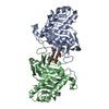
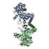
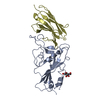
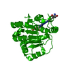
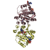
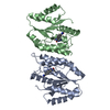
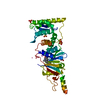
| Title | Pseudomonas aeruginosa HasA mutant - H32A | ||||||
 Components Components | HasAp | ||||||
 Keywords Keywords | METAL BINDING PROTEIN / HasA / hemophore | ||||||
| Function / homology | Haem-binding HasA / Haem-binding HasA superfamily / Heme-binding protein A (HasA) / metal ion binding / PROTOPORPHYRIN IX CONTAINING FE / HasAp Function and homology information Function and homology information | ||||||
| Biological species |   Pseudomonas aeruginosa (bacteria) Pseudomonas aeruginosa (bacteria) | ||||||
| Method |  X-RAY DIFFRACTION / SYNCHROTRON / MOLECULAR REPLACEMENT / Resolution: 2.096 Å X-RAY DIFFRACTION / SYNCHROTRON / MOLECULAR REPLACEMENT / Resolution: 2.096 Å | ||||||
 Authors Authors | Brimberry, M. / Lanzilotta, W. / Wilks, A. / Dent, A. | ||||||
| Funding support |  United States, 1items United States, 1items
| ||||||
 Citation Citation | Journal: To Be Published Title: Crystal Structure of Pseudomonas aeruginosa HasA mutant H32A Authors: Brimberry, M. / Lanzilotta, W. | ||||||
| History |
|
- Structure visualization
| Structure viewer | Molecule: MolmilJmol/JSmol |
|---|
- Downloads & links
-Download
| PDBx/mmCIF format | 6urb.cif.gz | 85.9 KB | Display | PDBx/mmCIF format |
|---|---|---|---|---|
| PDB format | pdb6urb.ent.gz | 63 KB | Display | PDB format |
| PDBx/mmJSON format | 6urb.json.gz | Tree view | PDBx/mmJSON format | |
| Others |  Other downloads Other downloads |
-Validation report
| Arichive directory | https://data.pdbj.org/pub/pdb/validation_reports/ur/6urbftp://data.pdbj.org/pub/pdb/validation_reports/ur/6urb | HTTPS FTP |
|---|
-Related structure data
| Related structure data | 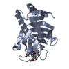 3ellS S: Starting model for refinement |
|---|---|
| Similar structure data |


-Links
 PDBj
PDBj- Assembly
| Deposited unit | 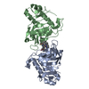
| ||||||||
|---|---|---|---|---|---|---|---|---|---|
| 1 |
| ||||||||
| Unit cell |
| ||||||||
| Components on special symmetry positions |
|
-Components
| #1: Protein | Mass: 20408.195 Da / Num. of mol.: 2 / Mutation: H32A Source method: isolated from a genetically manipulated source Source: (gene. exp.) Pseudomonas aeruginosa (bacteria) / Gene: hasAp, IPC3_06435 / Production host: #2: Chemical |   Mass: 616.487 Da / Num. of mol.: 2 / Source method: obtained synthetically / Formula: C34H32FeN4O4 / Feature type: SUBJECT OF INVESTIGATION Mass: 616.487 Da / Num. of mol.: 2 / Source method: obtained synthetically / Formula: C34H32FeN4O4 / Feature type: SUBJECT OF INVESTIGATION#3: Water | ChemComp-HOH / |  Mass: 18.015 Da / Num. of mol.: 90 / Source method: isolated from a natural source / Formula: H2O Mass: 18.015 Da / Num. of mol.: 90 / Source method: isolated from a natural source / Formula: H2OHas ligand of interest | Y | |
|---|
-Experimental details
-Experiment
| Experiment | Method: X-RAY DIFFRACTION / Number of used crystals: 1 |
|---|
- Sample preparation
| Crystal | Density Matthews: 1.95 Å3/Da / Density % sol: 37.03 % |
|---|---|
| Crystal grow | Temperature: 295 K / Method: vapor diffusion, sitting drop Details: 1M Sodium Citrate, 0.1M Tris HCl pH 7.4, 0.2M Sodium Chloride |
-Data collection
| Diffraction | Mean temperature: 291 K / Serial crystal experiment: N |
|---|---|
| Diffraction source | Source: SYNCHROTRON / Site: APS / Beamline: 22-ID / Wavelength: 1 Å |
| Detector | Type: MARMOSAIC 225 mm CCD / Detector: CCD / Date: Jul 25, 2019 |
| Radiation | Protocol: SINGLE WAVELENGTH / Monochromatic (M) / Laue (L): M / Scattering type: x-ray |
| Radiation wavelength | Wavelength: 1 Å / Relative weight: 1 |
| Reflection | Resolution: 2.09→50 Å / Num. obs: 17648 / % possible obs: 94 % / Redundancy: 3.6 % / CC1/2: 0.995 / Net I/σ(I): 17 |
| Reflection shell | Resolution: 2.09→2.17 Å / Mean I/σ(I) obs: 2.9 / Num. unique obs: 17648 / CC1/2: 0.91 |
- Processing
| Software |
| ||||||||||||||||||||||||||||||||||||||||||
|---|---|---|---|---|---|---|---|---|---|---|---|---|---|---|---|---|---|---|---|---|---|---|---|---|---|---|---|---|---|---|---|---|---|---|---|---|---|---|---|---|---|---|---|
| Refinement | Method to determine structure: MOLECULAR REPLACEMENT Starting model: 3ELL Resolution: 2.096→29.891 Å / SU ML: 0.26 / Cross valid method: THROUGHOUT / σ(F): 1.36 / Phase error: 31.33
| ||||||||||||||||||||||||||||||||||||||||||
| Solvent computation | Shrinkage radii: 0.9 Å / VDW probe radii: 1.11 Å | ||||||||||||||||||||||||||||||||||||||||||
| Displacement parameters | Biso max: 126.75 Å2 / Biso mean: 49.9748 Å2 / Biso min: 10.59 Å2 | ||||||||||||||||||||||||||||||||||||||||||
| Refinement step | Cycle: final / Resolution: 2.096→29.891 Å
| ||||||||||||||||||||||||||||||||||||||||||
| Refine LS restraints |
| ||||||||||||||||||||||||||||||||||||||||||
| LS refinement shell | Refine-ID: X-RAY DIFFRACTION / Rfactor Rfree error: 0
|
