 Movie
Movie Controller


+ Open data

- Basic information
| Entry | Database: PDB / ID: 2ox1 | ||||||
|---|---|---|---|---|---|---|---|
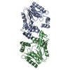
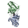
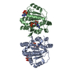
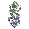
| Title | Archaeal Dehydroquinase | ||||||
 Components Components | 3-dehydroquinate dehydratase | ||||||
 Keywords Keywords | LYASE / (beta-alpha)8 barrel | ||||||
| Function / homology |  Function and homology information Function and homology information3,4-dihydroxybenzoate biosynthetic process / 3-dehydroquinate dehydratase / 3-dehydroquinate dehydratase activity / chorismate biosynthetic process / aromatic amino acid biosynthetic process / amino acid biosynthetic process Similarity search - Function | ||||||
| Biological species |   Archaeoglobus fulgidus (archaea) Archaeoglobus fulgidus (archaea) | ||||||
| Method |  X-RAY DIFFRACTION / SIRAS / Resolution: 2.33 Å X-RAY DIFFRACTION / SIRAS / Resolution: 2.33 Å | ||||||
 Authors Authors | Gallagher, D.T. / Smith, N.N. | ||||||
 Citation Citation | Journal: Acta Crystallogr.,Sect.F / Year: 2008 Title: Structure and lability of archaeal dehydroquinase. Authors: Smith, N.N. / Gallagher, D.T. | ||||||
| History |
|
- Structure visualization
| Structure viewer | Molecule: MolmilJmol/JSmol |
|---|
- Downloads & links
-Download
| PDBx/mmCIF format | 2ox1.cif.gz | 167 KB | Display | PDBx/mmCIF format |
|---|---|---|---|---|
| PDB format | pdb2ox1.ent.gz | 135.1 KB | Display | PDB format |
| PDBx/mmJSON format | 2ox1.json.gz | Tree view | PDBx/mmJSON format | |
| Others |  Other downloads Other downloads |
-Validation report
| Arichive directory | https://data.pdbj.org/pub/pdb/validation_reports/ox/2ox1ftp://data.pdbj.org/pub/pdb/validation_reports/ox/2ox1 | HTTPS FTP |
|---|
-Related structure data
| Similar structure data |
|---|


-Links
 PDBj
PDBj
- Assembly
| Deposited unit | 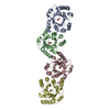
| ||||||||
|---|---|---|---|---|---|---|---|---|---|
| 1 | 
| ||||||||
| 2 | 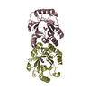
| ||||||||
| Unit cell |
| ||||||||
| Details | The biological unit is a dimer. The crystal asymmetric unit contains two biological dimers. |
-Components
| #1: Protein | Mass: 22237.307 Da / Num. of mol.: 4 Source method: isolated from a genetically manipulated source Source: (gene. exp.) Archaeoglobus fulgidus (archaea) / Gene: aroD / Plasmid: pET3a / Species (production host): Escherichia coli / Production host:  #2: Chemical | ChemComp-GOL /   Mass: 92.094 Da / Num. of mol.: 4 / Source method: obtained synthetically / Formula: C3H8O3 Mass: 92.094 Da / Num. of mol.: 4 / Source method: obtained synthetically / Formula: C3H8O3#3: Water | ChemComp-HOH / |  Mass: 18.015 Da / Num. of mol.: 385 / Source method: isolated from a natural source / Formula: H2O Mass: 18.015 Da / Num. of mol.: 385 / Source method: isolated from a natural source / Formula: H2O |
|---|
-Experimental details
-Experiment
| Experiment | Method: X-RAY DIFFRACTION / Number of used crystals: 1 |
|---|
- Sample preparation
| Crystal | Density Matthews: 2.91 Å3/Da / Density % sol: 57.68 % |
|---|---|
| Crystal grow | Temperature: 278 K / Method: vapor diffusion, hanging drop / pH: 4.6 Details: 0.2M ammonium acetate 0.1M sodium acetate 30% PEG 4000, pH 4.6, VAPOR DIFFUSION, HANGING DROP, temperature 278K |
-Data collection
| Diffraction | Mean temperature: 105 K |
|---|---|
| Diffraction source | Source: ROTATING ANODE / Type: RIGAKU / Wavelength: 1.5418 Å |
| Detector | Type: RIGAKU RAXIS IV / Detector: IMAGE PLATE / Date: Jan 31, 2004 / Details: mirrors |
| Radiation | Monochromator: mirror coating / Protocol: SINGLE WAVELENGTH / Monochromatic (M) / Laue (L): M / Scattering type: x-ray |
| Radiation wavelength | Wavelength: 1.5418 Å / Relative weight: 1 |
| Reflection | Resolution: 2.33→20 Å / Num. all: 43264 / Num. obs: 43264 / % possible obs: 96.2 % / Observed criterion σ(F): 0 / Observed criterion σ(I): 0 / Redundancy: 3.36 % / Rmerge(I) obs: 0.04 / Net I/σ(I): 17.7 |
| Reflection shell | Resolution: 2.33→2.41 Å / Redundancy: 3.27 % / Rmerge(I) obs: 0.293 / Mean I/σ(I) obs: 4 / Num. unique all: 4135 / % possible all: 93.3 |
- Processing
| Software |
| ||||||||||||||||||||||||||||||||||||||||||||||||||||||||||||||||||||||||||||||||||||||||||
|---|---|---|---|---|---|---|---|---|---|---|---|---|---|---|---|---|---|---|---|---|---|---|---|---|---|---|---|---|---|---|---|---|---|---|---|---|---|---|---|---|---|---|---|---|---|---|---|---|---|---|---|---|---|---|---|---|---|---|---|---|---|---|---|---|---|---|---|---|---|---|---|---|---|---|---|---|---|---|---|---|---|---|---|---|---|---|---|---|---|---|---|
| Refinement | Method to determine structure: SIRAS / Resolution: 2.33→10 Å / Cor.coef. Fo:Fc: 0.95 / Cor.coef. Fo:Fc free: 0.916 / SU B: 16.22 / SU ML: 0.201 / Cross valid method: THROUGHOUT / σ(F): 0 / ESU R: 0.343 / ESU R Free: 0.271 / Stereochemistry target values: MAXIMUM LIKELIHOOD / Details: HYDROGENS HAVE BEEN ADDED IN THE RIDING POSITIONS
| ||||||||||||||||||||||||||||||||||||||||||||||||||||||||||||||||||||||||||||||||||||||||||
| Solvent computation | Ion probe radii: 0.8 Å / Shrinkage radii: 0.8 Å / VDW probe radii: 1.2 Å / Solvent model: MASK | ||||||||||||||||||||||||||||||||||||||||||||||||||||||||||||||||||||||||||||||||||||||||||
| Displacement parameters | Biso mean: 30.226 Å2
| ||||||||||||||||||||||||||||||||||||||||||||||||||||||||||||||||||||||||||||||||||||||||||
| Refine analyze |
| ||||||||||||||||||||||||||||||||||||||||||||||||||||||||||||||||||||||||||||||||||||||||||
| Refinement step | Cycle: LAST / Resolution: 2.33→10 Å
| ||||||||||||||||||||||||||||||||||||||||||||||||||||||||||||||||||||||||||||||||||||||||||
| Refine LS restraints |
| ||||||||||||||||||||||||||||||||||||||||||||||||||||||||||||||||||||||||||||||||||||||||||
| LS refinement shell | Resolution: 2.33→2.387 Å / Total num. of bins used: 20
|
