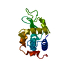
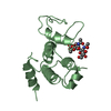
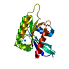
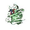
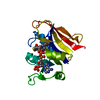
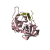
Mass: 16741.719 Da / Num. of mol.: 1 Source method: isolated from a genetically manipulated source Source: (gene. exp.) Penaeus vannamei (Pacific white shrimp) Production host: Escherichia coli (E. coli) / References: UniProt: Q95V66, lysozyme
Resolution: 2.25→35.692 Å / Cor.coef. Fo:Fc: 0.935 / Cor.coef. Fo:Fc free: 0.901 / SU B: 6.762 / SU ML: 0.161 / Cross valid method: THROUGHOUT / ESU R: 0.52 / ESU R Free: 0.273 Details: Hydrogens have been added in their riding positions
Rfactor
Num. reflection
% reflection
Rfree
0.2463
622
9.92 %
Rwork
0.1998
-
-
all
0.204
-
-
obs
-
6270
86.162 %
Solvent computation
Ion probe radii: 0.8 Å / Shrinkage radii: 0.8 Å / VDW probe radii: 1.2 Å
Displacement parameters
Biso mean: 23.065 Å2
Baniso -1
Baniso -2
Baniso -3
1-
-0.268 Å2
-0.134 Å2
0 Å2
2-
-
-0.268 Å2
0 Å2
3-
-
-
0.871 Å2
Refinement step
Cycle: LAST / Resolution: 2.25→35.692 Å
Protein
Nucleic acid
Ligand
Solvent
Total
Num. atoms
1084
0
6
66
1156
Refine LS restraints
Refine-ID
Type
Dev ideal
Dev ideal target
Number
X-RAY DIFFRACTION
r_bond_refined_d
0.008
0.013
1115
X-RAY DIFFRACTION
r_bond_other_d
0.001
0.018
980
X-RAY DIFFRACTION
r_ext_dist_refined_d
0.198
0.01
82
X-RAY DIFFRACTION
r_angle_refined_deg
1.527
1.646
1500
X-RAY DIFFRACTION
r_angle_other_deg
1.382
1.594
2268
X-RAY DIFFRACTION
r_dihedral_angle_1_deg
7.681
5
132
X-RAY DIFFRACTION
r_dihedral_angle_2_deg
32.047
21.389
72
X-RAY DIFFRACTION
r_dihedral_angle_3_deg
15.192
15
194
X-RAY DIFFRACTION
r_dihedral_angle_4_deg
22.238
15
11
X-RAY DIFFRACTION
r_chiral_restr
0.068
0.2
139
X-RAY DIFFRACTION
r_gen_planes_refined
0.007
0.02
1265
X-RAY DIFFRACTION
r_gen_planes_other
0.001
0.02
272
X-RAY DIFFRACTION
r_nbd_refined
0.213
0.2
221
X-RAY DIFFRACTION
r_symmetry_nbd_other
0.195
0.2
935
X-RAY DIFFRACTION
r_nbtor_refined
0.174
0.2
545
X-RAY DIFFRACTION
r_symmetry_nbtor_other
0.076
0.2
490
X-RAY DIFFRACTION
r_xyhbond_nbd_refined
0.181
0.2
60
X-RAY DIFFRACTION
r_symmetry_xyhbond_nbd_other
0.068
0.2
1
X-RAY DIFFRACTION
r_symmetry_nbd_refined
0.025
0.2
5
X-RAY DIFFRACTION
r_nbd_other
0.186
0.2
32
X-RAY DIFFRACTION
r_symmetry_xyhbond_nbd_refined
0.11
0.2
4
X-RAY DIFFRACTION
r_mcbond_it
1.66
2.314
531
X-RAY DIFFRACTION
r_mcbond_other
1.659
2.31
530
X-RAY DIFFRACTION
r_mcangle_it
2.614
3.461
662
X-RAY DIFFRACTION
r_mcangle_other
2.612
3.465
663
X-RAY DIFFRACTION
r_scbond_it
1.995
2.549
584
X-RAY DIFFRACTION
r_scbond_other
1.993
2.553
585
X-RAY DIFFRACTION
r_scangle_it
3.16
3.714
838
X-RAY DIFFRACTION
r_scangle_other
3.158
3.718
839
X-RAY DIFFRACTION
r_lrange_it
5.68
42.407
4694
X-RAY DIFFRACTION
r_lrange_other
5.654
42.39
4660
LS refinement shell
Resolution (Å)
Rfactor Rfree
Num. reflection Rfree
Rfactor Rwork
Num. reflection Rwork
Refine-ID
% reflection obs (%)
2.25-2.309
0.392
46
0.286
445
X-RAY DIFFRACTION
89.7623
2.309-2.372
0.261
46
0.254
418
X-RAY DIFFRACTION
91.5187
2.372-2.441
0.262
46
0.224
407
X-RAY DIFFRACTION
89.881
2.441-2.516
0.244
46
0.22
417
X-RAY DIFFRACTION
92.9719
2.516-2.598
0.334
37
0.212
392
X-RAY DIFFRACTION
88.4536
2.598-2.689
0.248
41
0.207
351
X-RAY DIFFRACTION
86.918
2.689-2.791
0.267
39
0.199
347
X-RAY DIFFRACTION
87.7273
2.791-2.904
0.281
34
0.214
336
X-RAY DIFFRACTION
87.6777
2.904-3.033
0.261
39
0.187
325
X-RAY DIFFRACTION
86.052
3.033-3.181
0.208
34
0.185
300
X-RAY DIFFRACTION
88.1266
3.181-3.353
0.212
32
0.187
298
X-RAY DIFFRACTION
87.766
3.353-3.556
0.202
35
0.188
260
X-RAY DIFFRACTION
81.9444
3.556-3.8
0.252
23
0.176
241
X-RAY DIFFRACTION
78.806
3.8-4.104
0.193
24
0.179
216
X-RAY DIFFRACTION
77.4193
4.104-4.494
0.277
22
0.162
205
X-RAY DIFFRACTION
78.8194
4.494-5.022
0.176
23
0.164
196
X-RAY DIFFRACTION
83.27
5.794-7.085
0.407
18
0.212
155
X-RAY DIFFRACTION
83.9806
+
About Yorodumi
-
News
-
Feb 9, 2022. New format data for meta-information of EMDB entries
New format data for meta-information of EMDB entries
Version 3 of the EMDB header file is now the official format.
The previous official version 1.9 will be removed from the archive.
In the structure databanks used in Yorodumi, some data are registered as the other names, "COVID-19 virus" and "2019-nCoV". Here are the details of the virus and the list of structure data.
Jan 31, 2019. EMDB accession codes are about to change! (news from PDBe EMDB page)
EMDB accession codes are about to change! (news from PDBe EMDB page)
The allocation of 4 digits for EMDB accession codes will soon come to an end. Whilst these codes will remain in use, new EMDB accession codes will include an additional digit and will expand incrementally as the available range of codes is exhausted. The current 4-digit format prefixed with “EMD-” (i.e. EMD-XXXX) will advance to a 5-digit format (i.e. EMD-XXXXX), and so on. It is currently estimated that the 4-digit codes will be depleted around Spring 2019, at which point the 5-digit format will come into force.
The EM Navigator/Yorodumi systems omit the EMD- prefix.
Related info.:Q: What is EMD? / ID/Accession-code notation in Yorodumi/EM Navigator
Yorodumi is a browser for structure data from EMDB, PDB, SASBDB, etc.
This page is also the successor to EM Navigator detail page, and also detail information page/front-end page for Omokage search.
The word "yorodu" (or yorozu) is an old Japanese word meaning "ten thousand". "mi" (miru) is to see.
Related info.:EMDB / PDB / SASBDB / Comparison of 3 databanks / Yorodumi Search / Aug 31, 2016. New EM Navigator & Yorodumi / Yorodumi Papers / Jmol/JSmol / Function and homology information / Changes in new EM Navigator and Yorodumi
 Movie
Movie Controller


 Open data
 Basic information
 Components
 Keywords
 Function and homology information
 Penaeus vannamei (Pacific white shrimp)
 X-RAY DIFFRACTION /
  Authors
 Citation
 Structure visualization
 Downloads & links
 Other downloads


 PDBj
PDBj
 Assembly


 Mass: 92.094 Da / Num. of mol.: 1 / Source method: obtained synthetically / Formula: C3H8O3 / Feature type: SUBJECT OF INVESTIGATION
 Mass: 18.015 Da / Num. of mol.: 66 / Source method: isolated from a natural source / Formula: H2O
 Sample preparation
 Processing