Resolution: 1.92→19.72 Å / Cor.coef. Fo:Fc: 0.935 / Cor.coef. Fo:Fc free: 0.902 / WRfactor Rfree: 0.239 / WRfactor Rwork: 0.2 / SU B: 4.496 / SU ML: 0.131 / Cross valid method: THROUGHOUT / σ(F): 0 / ESU R: 0.193 / ESU R Free: 0.177 / Stereochemistry target values: MAXIMUM LIKELIHOOD Details: HYDROGENS HAVE BEEN ADDED IN THE RIDING POSITIONS; U VALUES: REFINED INDIVIDUALLY. ARP/WARP, COOT and MOLPROBITY were also used during refinement
Rfactor
Num. reflection
% reflection
Selection details
Rfree
0.278
682
5.077 %
RANDOM
Rwork
0.23
-
-
-
obs
0.233
13432
99.918 %
-
Solvent computation
Ion probe radii: 0.8 Å / Shrinkage radii: 0.8 Å / VDW probe radii: 1.4 Å / Solvent model: MASK BULK SOLVENT
Displacement parameters
Biso mean: 18.599 Å2
Baniso -1
Baniso -2
Baniso -3
1-
-1.489 Å2
0 Å2
0 Å2
2-
-
0.299 Å2
0 Å2
3-
-
-
1.19 Å2
Refinement step
Cycle: LAST / Resolution: 1.92→19.72 Å
Protein
Nucleic acid
Ligand
Solvent
Total
Num. atoms
1297
0
3
50
1350
Refine LS restraints
Refine-ID
Type
Dev ideal
Dev ideal target
Number
X-RAY DIFFRACTION
r_bond_refined_d
0.016
0.022
1326
X-RAY DIFFRACTION
r_bond_other_d
0.005
0.02
874
X-RAY DIFFRACTION
r_angle_refined_deg
1.498
1.963
1810
X-RAY DIFFRACTION
r_angle_other_deg
1.007
3
2139
X-RAY DIFFRACTION
r_dihedral_angle_1_deg
6.477
5
176
X-RAY DIFFRACTION
r_dihedral_angle_2_deg
32.686
24.138
58
X-RAY DIFFRACTION
r_dihedral_angle_3_deg
13.402
15
214
X-RAY DIFFRACTION
r_dihedral_angle_4_deg
20.293
15
8
X-RAY DIFFRACTION
r_chiral_restr
0.096
0.2
210
X-RAY DIFFRACTION
r_gen_planes_refined
0.006
0.02
1507
X-RAY DIFFRACTION
r_gen_planes_other
0.001
0.02
276
X-RAY DIFFRACTION
r_mcbond_it
0.897
1.5
843
X-RAY DIFFRACTION
r_mcbond_other
0.249
1.5
344
X-RAY DIFFRACTION
r_mcangle_it
1.57
2
1362
X-RAY DIFFRACTION
r_scbond_it
2.309
3
483
X-RAY DIFFRACTION
r_scangle_it
3.648
4.5
443
LS refinement shell
Refine-ID: X-RAY DIFFRACTION / Total num. of bins used: 20
Resolution (Å)
Rfactor Rfree
Num. reflection Rfree
Rfactor Rwork
Num. reflection Rwork
Num. reflection all
% reflection obs (%)
1.92-1.969
0.474
48
0.35
914
963
99.896
1.969-2.023
0.415
46
0.312
907
953
100
2.023-2.081
0.311
45
0.282
875
921
99.891
2.081-2.144
0.329
45
0.263
834
882
99.66
2.144-2.214
0.299
42
0.238
813
855
100
2.214-2.29
0.463
31
0.347
820
857
99.3
2.29-2.375
0.258
48
0.222
763
811
100
2.375-2.471
0.32
37
0.226
749
786
100
2.471-2.579
0.316
43
0.225
707
750
100
2.579-2.703
0.26
40
0.231
689
729
100
2.703-2.846
0.275
38
0.237
652
690
100
2.846-3.014
0.323
38
0.231
617
655
100
3.014-3.217
0.212
33
0.216
579
612
100
3.217-3.467
0.262
29
0.218
560
589
100
3.467-3.787
0.238
29
0.21
507
536
100
3.787-4.214
0.254
30
0.189
468
498
100
4.214-4.829
0.135
18
0.157
424
442
100
4.829-5.828
0.184
18
0.212
362
380
100
5.828-7.904
0.297
17
0.248
302
319
100
7.904-19.72
0.449
7
0.226
208
215
100
+
About Yorodumi
-
News
-
Feb 9, 2022. New format data for meta-information of EMDB entries
New format data for meta-information of EMDB entries
Version 3 of the EMDB header file is now the official format.
The previous official version 1.9 will be removed from the archive.
In the structure databanks used in Yorodumi, some data are registered as the other names, "COVID-19 virus" and "2019-nCoV". Here are the details of the virus and the list of structure data.
Jan 31, 2019. EMDB accession codes are about to change! (news from PDBe EMDB page)
EMDB accession codes are about to change! (news from PDBe EMDB page)
The allocation of 4 digits for EMDB accession codes will soon come to an end. Whilst these codes will remain in use, new EMDB accession codes will include an additional digit and will expand incrementally as the available range of codes is exhausted. The current 4-digit format prefixed with “EMD-” (i.e. EMD-XXXX) will advance to a 5-digit format (i.e. EMD-XXXXX), and so on. It is currently estimated that the 4-digit codes will be depleted around Spring 2019, at which point the 5-digit format will come into force.
The EM Navigator/Yorodumi systems omit the EMD- prefix.
Related info.:Q: What is EMD? / ID/Accession-code notation in Yorodumi/EM Navigator
Yorodumi is a browser for structure data from EMDB, PDB, SASBDB, etc.
This page is also the successor to EM Navigator detail page, and also detail information page/front-end page for Omokage search.
The word "yorodu" (or yorozu) is an old Japanese word meaning "ten thousand". "mi" (miru) is to see.
Related info.:EMDB / PDB / SASBDB / Comparison of 3 databanks / Yorodumi Search / Aug 31, 2016. New EM Navigator & Yorodumi / Yorodumi Papers / Jmol/JSmol / Function and homology information / Changes in new EM Navigator and Yorodumi
 Movie
Movie Controller


 Open data
 Basic information
 Components
 Keywords
 Function and homology information
 Homo sapiens (human)
 X-RAY DIFFRACTION /
  Authors
 Citation
 Structure visualization
 Downloads & links
 Other downloads
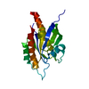


 PDBj
PDBj Assembly
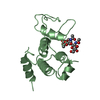
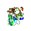
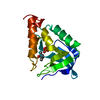
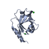
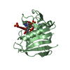
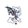
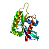


 Num. of mol.: 3 / Source method: obtained synthetically
 Mass: 18.015 Da / Num. of mol.: 50 / Source method: isolated from a natural source / Formula: H2O
 Sample preparation
 Processing