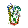
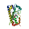
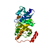
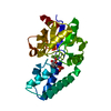
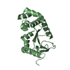
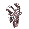
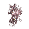
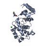
[English] 日本語
Yorodumi
- PDB-6uie: Structure of the cytoplasmic domain of the T3SS sorting platform ... -

+
Open data


ID or keywords:

Loading...

-
Basic information

Entry
Database: PDB / ID: 6uie
TitleStructure of the cytoplasmic domain of the T3SS sorting platform protein PscK from P. aeruginosa
ComponentsType III export protein PscK
KeywordsTRANSPORT PROTEIN / PscK / type III secretion apparatus protein
Function / homologyType III secretion system, secretion protein K / YOP proteins translocation protein K (YscK) / protein secretion by the type III secretion system / : / Type III export protein PscK
Function and homology information
Biological speciesPseudomonas aeruginosa (bacteria)
MethodX-RAY DIFFRACTION / SYNCHROTRON / SAD / Resolution: 2.55 Å
AuthorsMuthuramalingam, M. / Lovell, S. / Battaile, K.P. / Picking, W.D.
Funding support United States, 3items
OrganizationGrant numberCountry
National Institutes of Health/National Institute Of Allergy and Infectious Diseases (NIH/NIAID)R01 AI123351 United States
National Institutes of Health/National Institute Of Allergy and Infectious Diseases (NIH/NIAID)R21 AI146517 United States
National Institutes of Health/National Institute of General Medical Sciences (NIH/NIGMS)P30 GM110761 United States
CitationJournal: J.Mol.Biol. / Year: 2020
Title: The Structures of SctK and SctD from Pseudomonas aeruginosa Reveal the Interface of the Type III Secretion System Basal Body and Sorting Platform.
Authors: Muthuramalingam, M. / Whittier, S.K. / Lovell, S. / Battaile, K.P. / Tachiyama, S. / Johnson, D.K. / Picking, W.L. / Picking, W.D.
History
DepositionSep 30, 2019Deposition site: RCSB / Processing site: RCSB
Revision 1.0Oct 7, 2020Provider: repository / Type: Initial release
Revision 1.1Nov 4, 2020Group: Database references / Category: citation / citation_author
Item: _citation.country / _citation.journal_abbrev ..._citation.country / _citation.journal_abbrev / _citation.journal_id_ASTM / _citation.journal_id_CSD / _citation.journal_id_ISSN / _citation.pdbx_database_id_DOI / _citation.title / _citation.year
Revision 1.2Nov 11, 2020Group: Database references / Category: citation / Item: _citation.pdbx_database_id_PubMed / _citation.title
Revision 1.3Dec 16, 2020Group: Database references / Category: citation
Item: _citation.journal_volume / _citation.page_first / _citation.page_last
Revision 1.4Oct 11, 2023Group: Data collection / Database references / Refinement description
Category: chem_comp_atom / chem_comp_bond ...chem_comp_atom / chem_comp_bond / database_2 / pdbx_initial_refinement_model
Item: _database_2.pdbx_DOI / _database_2.pdbx_database_accession
Revision 1.5Nov 15, 2023Group: Data collection / Category: chem_comp_atom / chem_comp_bond / Item: _chem_comp_atom.atom_id / _chem_comp_bond.atom_id_2

-
Structure visualization

Structure viewerMolecule:
MolmilJmol/JSmol

Downloads & links

-
Assembly

Deposited unit
A: Type III export protein PscK
B: Type III export protein PscK
hetero molecules


Theoretical massNumber of molelcules
Total (without water)48,4653
Polymers48,4292
Non-polymers351
Water0
1
A: Type III export protein PscK
hetero molecules


Theoretical massNumber of molelcules
Total (without water)24,2502
Polymers24,2151
Non-polymers351
Water0
TypeNameSymmetry operationNumber
identity operation1_555x,y,z1
MethodPISA
2
B: Type III export protein PscK


Theoretical massNumber of molelcules
Total (without water)24,2151
Polymers24,2151
Non-polymers00
Water0
TypeNameSymmetry operationNumber
identity operation1_555x,y,z1
MethodPISA
Unit cell
Length a, b, c (Å)56.722, 81.733, 96.520
Angle α, β, γ (deg.)90.000, 90.000, 90.000
Int Tables number19
Space group name H-MP212121

-
Components

#1: Protein Type III export protein PscK


Mass: 24214.645 Da / Num. of mol.: 2
Source method: isolated from a genetically manipulated source
Source: (gene. exp.) Pseudomonas aeruginosa (bacteria) / Gene: pscK / Plasmid: pT7HMT / Production host: Escherichia coli (E. coli) / Strain (production host): Tuner (DE3) / References: UniProt: Q9I313
#2: Chemical ChemComp-CL / CHLORIDE ION / Chloride


Mass: 35.453 Da / Num. of mol.: 1 / Source method: obtained synthetically / Formula: Cl
Has ligand of interestY

-
Experimental details

-
Experiment

ExperimentMethod: X-RAY DIFFRACTION / Number of used crystals: 1

-
Sample preparation

CrystalDensity Matthews: 2.32 Å3/Da / Density % sol: 47.06 % / Mosaicity: 0 °
Crystal growTemperature: 291 K / Method: vapor diffusion, sitting drop / pH: 7.5
Details: 20% (w/v) PEG 1500, 100 mM Tris, 100 mM ammonium sulfate

-
Data collection

DiffractionMean temperature: 100 K / Serial crystal experiment: N
Diffraction sourceSource: SYNCHROTRON / Site: APS / Beamline: 17-ID / Wavelength: 0.97918 Å
DetectorType: DECTRIS PILATUS 6M / Detector: PIXEL / Date: Oct 13, 2018
RadiationProtocol: SINGLE WAVELENGTH / Monochromatic (M) / Laue (L): M / Scattering type: x-ray
Radiation wavelengthWavelength: 0.97918 Å / Relative weight: 1
ReflectionResolution: 2.55→48.9 Å / Num. obs: 28173 / % possible obs: 100 % / Redundancy: 63 % / CC1/2: 0.999 / Rmerge(I) obs: 0.26 / Net I/σ(I): 15
Reflection shell

Diffraction-ID: 1

Resolution (Å)Redundancy (%)Rmerge(I) obsNum. measured allNum. unique obsCC1/2Net I/σ(I) obs% possible all
2.55-2.6664.42.00811734718230.9763.3100
8.83-48.956.20.093244614350.9993599.8

-
Phasing

PhasingMethod: SAD

-
Processing

Software
NameVersionClassification
PHENIX1.17rc2_3615refinement
XDSdata reduction
Aimless0.7.4data scaling
PDB_EXTRACT3.25data extraction
CRANK2phasing
RefinementMethod to determine structure: SAD
Starting model: 4A0E
Resolution: 2.55→48.9 Å / SU ML: 0.37 / Cross valid method: THROUGHOUT / σ(F): 1.91 / Phase error: 36.6 / Stereochemistry target values: ML
RfactorNum. reflection% reflection
Rfree0.2936 1329 4.72 %
Rwork0.2359 26844 -
obs0.2388 28173 99.69 %
Solvent computationShrinkage radii: 0.9 Å / VDW probe radii: 1.11 Å / Solvent model: FLAT BULK SOLVENT MODEL
Displacement parametersBiso max: 123.45 Å2 / Biso mean: 61.4759 Å2 / Biso min: 28.66 Å2
Refinement stepCycle: final / Resolution: 2.55→48.9 Å
ProteinNucleic acidLigandSolventTotal
Num. atoms2745 0 1 0 2746
Biso mean--71.2 --
Num. residues----366
LS refinement shell

Refine-ID: X-RAY DIFFRACTION / Rfactor Rfree error: 0 / Total num. of bins used: 9

Resolution (Å)Rfactor RfreeNum. reflection RfreeRfactor RworkNum. reflection RworkNum. reflection all% reflection obs (%)
2.55-2.650.40911450.31882979312499
2.65-2.770.32951740.26682926310099
2.77-2.920.40551380.273430113149100
2.92-3.10.30171580.263929903148100
3.1-3.340.36961090.254330013110100
3.34-3.680.36911460.23729813127100
3.68-4.210.24711450.218129933138100
4.21-5.30.23651480.207629983146100
5.3-48.90.26651660.2329653131100

+
About Yorodumi

-
News

-
Feb 9, 2022. New format data for meta-information of EMDB entries

New format data for meta-information of EMDB entries

  • Version 3 of the EMDB header file is now the official format.
  • The previous official version 1.9 will be removed from the archive.

Related info.:EMDB header

External links:wwPDB to switch to version 3 of the EMDB data model

-
Aug 12, 2020. Covid-19 info

Covid-19 info

URL: https://pdbj.org/emnavi/covid19.php

New page: Covid-19 featured information page in EM Navigator.

Related info.:Covid-19 info / Mar 5, 2020. Novel coronavirus structure data

+
Mar 5, 2020. Novel coronavirus structure data

Novel coronavirus structure data

Related info.:Yorodumi Speices / Aug 12, 2020. Covid-19 info

External links:COVID-19 featured content - PDBj / Molecule of the Month (242):Coronavirus Proteases

+
Jan 31, 2019. EMDB accession codes are about to change! (news from PDBe EMDB page)

EMDB accession codes are about to change! (news from PDBe EMDB page)

  • The allocation of 4 digits for EMDB accession codes will soon come to an end. Whilst these codes will remain in use, new EMDB accession codes will include an additional digit and will expand incrementally as the available range of codes is exhausted. The current 4-digit format prefixed with “EMD-” (i.e. EMD-XXXX) will advance to a 5-digit format (i.e. EMD-XXXXX), and so on. It is currently estimated that the 4-digit codes will be depleted around Spring 2019, at which point the 5-digit format will come into force.
  • The EM Navigator/Yorodumi systems omit the EMD- prefix.

Related info.:Q: What is EMD? / ID/Accession-code notation in Yorodumi/EM Navigator

External links:EMDB Accession Codes are Changing Soon! / Contact to PDBj

+
Jul 12, 2017. Major update of PDB

Major update of PDB

  • wwPDB released updated PDB data conforming to the new PDBx/mmCIF dictionary.
  • This is a major update changing the version number from 4 to 5, and with Remediation, in which all the entries are updated.
  • In this update, many items about electron microscopy experimental information are reorganized (e.g. em_software).
  • Now, EM Navigator and Yorodumi are based on the updated data.

External links:wwPDB Remediation / Enriched Model Files Conforming to OneDep Data Standards Now Available in the PDB FTP Archive

-
Yorodumi

Thousand views of thousand structures

  • Yorodumi is a browser for structure data from EMDB, PDB, SASBDB, etc.
  • This page is also the successor to EM Navigator detail page, and also detail information page/front-end page for Omokage search.
  • The word "yorodu" (or yorozu) is an old Japanese word meaning "ten thousand". "mi" (miru) is to see.

Related info.:EMDB / PDB / SASBDB / Comparison of 3 databanks / Yorodumi Search / Aug 31, 2016. New EM Navigator & Yorodumi / Yorodumi Papers / Jmol/JSmol / Function and homology information / Changes in new EM Navigator and Yorodumi

Read more