 Movie
Movie Controller

[English] 日本語
 Yorodumi
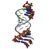
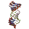
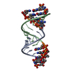
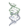
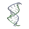
- PDB-6ugi: Crystal structure of a fragment of E. coli tRNA(Asp) consisting o... -
+ Open data

- Basic information
| Entry | Database: PDB / ID: 6ugi | |||||||||||||||
|---|---|---|---|---|---|---|---|---|---|---|---|---|---|---|---|---|
| Title | Crystal structure of a fragment of E. coli tRNA(Asp) consisting of its acceptor stem/T stem-loop. Long unit cell. | |||||||||||||||
 Components Components | tRNA(Asp) acceptor stem/T stem-loop | |||||||||||||||
 Keywords Keywords | RNA / tRNA RNA unmodified T-loop acceptor stem | |||||||||||||||
| Function / homology | RNA / RNA (> 10) Function and homology information Function and homology information | |||||||||||||||
| Biological species |  | |||||||||||||||
| Method |  X-RAY DIFFRACTION / SYNCHROTRON / MOLECULAR REPLACEMENT / Resolution: 1.75 Å X-RAY DIFFRACTION / SYNCHROTRON / MOLECULAR REPLACEMENT / Resolution: 1.75 Å | |||||||||||||||
 Authors Authors | Chan, C.W. / Mondragon, A. | |||||||||||||||
| Funding support |  United States, 4items United States, 4items
| |||||||||||||||
 Citation Citation | Journal: Rna / Year: 2020 Title: Crystal structures of an unmodified bacterial tRNA reveal intrinsic structural flexibility and plasticity as general properties of unbound tRNAs. Authors: Chan, C.W. / Badong, D. / Rajan, R. / Mondragon, A. | |||||||||||||||
| History |
|
- Structure visualization
| Structure viewer | Molecule: MolmilJmol/JSmol |
|---|
- Downloads & links
-Download
| PDBx/mmCIF format | 6ugi.cif.gz | 30.7 KB | Display | PDBx/mmCIF format |
|---|---|---|---|---|
| PDB format | pdb6ugi.ent.gz | 19.8 KB | Display | PDB format |
| PDBx/mmJSON format | 6ugi.json.gz | Tree view | PDBx/mmJSON format | |
| Others |  Other downloads Other downloads |
-Validation report
| Arichive directory | https://data.pdbj.org/pub/pdb/validation_reports/ug/6ugiftp://data.pdbj.org/pub/pdb/validation_reports/ug/6ugi | HTTPS FTP |
|---|
-Related structure data
| Related structure data |  6uggC  6ugjSC S: Starting model for refinement C: citing same article ( |
|---|---|
| Similar structure data |


-Links
 PDBj
PDBj


- Assembly
| Deposited unit | 
| |||||||||||||||||||||||||||||||||||||||
|---|---|---|---|---|---|---|---|---|---|---|---|---|---|---|---|---|---|---|---|---|---|---|---|---|---|---|---|---|---|---|---|---|---|---|---|---|---|---|---|---|
| 1 |
| |||||||||||||||||||||||||||||||||||||||
| Unit cell |
| |||||||||||||||||||||||||||||||||||||||
| Components on special symmetry positions |
|
-Components
| #1: RNA chain | Mass: 9989.942 Da / Num. of mol.: 1 / Source method: obtained synthetically / Source: (synth.) |
|---|---|
| #2: Chemical | ChemComp-SO4 /   Mass: 96.063 Da / Num. of mol.: 1 / Source method: obtained synthetically / Formula: SO4 Mass: 96.063 Da / Num. of mol.: 1 / Source method: obtained synthetically / Formula: SO4 |
| #3: Water | ChemComp-HOH /  Mass: 18.015 Da / Num. of mol.: 70 / Source method: isolated from a natural source / Formula: H2O Mass: 18.015 Da / Num. of mol.: 70 / Source method: isolated from a natural source / Formula: H2O |
| Has ligand of interest | N |
-Experimental details
-Experiment
| Experiment | Method: X-RAY DIFFRACTION / Number of used crystals: 1 |
|---|
- Sample preparation
| Crystal | Density Matthews: 2.57 Å3/Da / Density % sol: 52.15 % |
|---|---|
| Crystal grow | Temperature: 287 K / Method: vapor diffusion, hanging drop / Details: 2 M ammonium sulfate |
-Data collection
| Diffraction | Mean temperature: 100 K Ambient temp details: flash frozen with liquid nitrogen in crystallization well solution supplemented with either increased ammonium sulfate concentration or with 20% (v/v) glycerol for cryo-protection Serial crystal experiment: N |
|---|---|
| Diffraction source | Source: SYNCHROTRON / Site: APS / Beamline: 21-ID-D / Wavelength: 1.1272 Å |
| Detector | Type: DECTRIS EIGER X 9M / Detector: PIXEL / Date: Nov 15, 2018 / Details: Monochromator |
| Radiation | Monochromator: Si(111) / Protocol: SINGLE WAVELENGTH / Monochromatic (M) / Laue (L): M / Scattering type: x-ray |
| Radiation wavelength | Wavelength: 1.1272 Å / Relative weight: 1 |
| Reflection | Resolution: 1.75→37.38 Å / Num. obs: 9226 / % possible obs: 89 % / Redundancy: 8.1 % / CC1/2: 0.997 / Rmerge(I) obs: 0.118 / Rpim(I) all: 0.045 / Rrim(I) all: 0.126 / Net I/σ(I): 8.2 |
| Reflection shell | Resolution: 1.754→1.872 Å / Redundancy: 7.4 % / Rmerge(I) obs: 1.257 / Num. unique obs: 3401 / CC1/2: 0.683 / Rpim(I) all: 0.494 / Rrim(I) all: 1.354 / % possible all: 34.9 |
- Processing
| Software |
| ||||||||||||||||||||||||||||||||||||||||
|---|---|---|---|---|---|---|---|---|---|---|---|---|---|---|---|---|---|---|---|---|---|---|---|---|---|---|---|---|---|---|---|---|---|---|---|---|---|---|---|---|---|
| Refinement | Method to determine structure: MOLECULAR REPLACEMENT Starting model: PDB entry 6UGJ Resolution: 1.75→37.38 Å / Cor.coef. Fo:Fc: 0.948 / Cor.coef. Fo:Fc free: 0.942 / SU B: 3.066 / SU ML: 0.091 / Cross valid method: THROUGHOUT / σ(F): 0 / ESU R: 0.156 / ESU R Free: 0.141 Details: HYDROGENS HAVE BEEN ADDED IN THE RIDING POSITIONS U VALUES : REFINED INDIVIDUALLY
| ||||||||||||||||||||||||||||||||||||||||
| Solvent computation | Ion probe radii: 0.8 Å / Shrinkage radii: 0.8 Å / VDW probe radii: 1.2 Å | ||||||||||||||||||||||||||||||||||||||||
| Displacement parameters | Biso max: 126.84 Å2 / Biso mean: 33.009 Å2 / Biso min: 17.78 Å2
| ||||||||||||||||||||||||||||||||||||||||
| Refinement step | Cycle: final / Resolution: 1.75→37.38 Å
| ||||||||||||||||||||||||||||||||||||||||
| Refine LS restraints |
| ||||||||||||||||||||||||||||||||||||||||
| LS refinement shell | Resolution: 1.752→1.798 Å / Rfactor Rfree error: 0 / Total num. of bins used: 20
|
