 Movie
Movie Controller


+ Open data

- Basic information
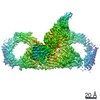
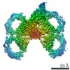
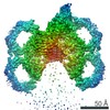
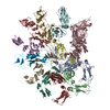
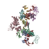
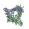
| Entry | Database: PDB / ID: 6uea | |||||||||
|---|---|---|---|---|---|---|---|---|---|---|
| Title | Structure of pentameric sIgA complex | |||||||||
 Components Components |
| |||||||||
 Keywords Keywords | IMMUNE SYSTEM / immunoglobulin | |||||||||
| Function / homology |  Function and homology information Function and homology informationpolymeric immunoglobulin receptor activity / immunoglobulin transcytosis in epithelial cells mediated by polymeric immunoglobulin receptor / polymeric immunoglobulin binding / dimeric IgA immunoglobulin complex / secretory dimeric IgA immunoglobulin complex / monomeric IgA immunoglobulin complex / secretory IgA immunoglobulin complex / pentameric IgM immunoglobulin complex / Fc receptor signaling pathway / IgA binding ...polymeric immunoglobulin receptor activity / immunoglobulin transcytosis in epithelial cells mediated by polymeric immunoglobulin receptor / polymeric immunoglobulin binding / dimeric IgA immunoglobulin complex / secretory dimeric IgA immunoglobulin complex / monomeric IgA immunoglobulin complex / secretory IgA immunoglobulin complex / pentameric IgM immunoglobulin complex / Fc receptor signaling pathway / IgA binding / IgA immunoglobulin complex / glomerular filtration / detection of chemical stimulus involved in sensory perception of bitter taste / immunoglobulin complex, circulating / immunoglobulin receptor binding / azurophil granule membrane / positive regulation of respiratory burst / receptor clustering / humoral immune response / complement activation, classical pathway / Scavenging of heme from plasma / antigen binding / Cell surface interactions at the vascular wall / B cell receptor signaling pathway / epidermal growth factor receptor signaling pathway / transmembrane signaling receptor activity / antibacterial humoral response / protein-containing complex assembly / blood microparticle / adaptive immune response / protein-macromolecule adaptor activity / signaling receptor complex / immune response / innate immune response / Neutrophil degranulation / signal transduction / protein homodimerization activity / : / extracellular exosome / extracellular region / plasma membrane Similarity search - Function | |||||||||
| Biological species |  Homo sapiens (human) Homo sapiens (human) | |||||||||
| Method | ELECTRON MICROSCOPY / single particle reconstruction / cryo EM / Resolution: 3 Å | |||||||||
 Authors Authors | Kumar, N. / Arthur, C.P. / Ciferri, C. / Matsumoto, M.L. | |||||||||
 Citation Citation | Journal: Science / Year: 2020 Title: Structure of the secretory immunoglobulin A core. Authors: Nikit Kumar / Christopher P Arthur / Claudio Ciferri / Marissa L Matsumoto /  Abstract: Secretory immunoglobulin A (sIgA) represents the immune system's first line of defense against mucosal pathogens. IgAs are transported across the epithelium, as dimers and higher-order polymers, by ...Secretory immunoglobulin A (sIgA) represents the immune system's first line of defense against mucosal pathogens. IgAs are transported across the epithelium, as dimers and higher-order polymers, by the polymeric immunoglobulin receptor (pIgR). Upon reaching the luminal side, sIgAs mediate host protection and pathogen neutralization. In recent years, an increasing amount of attention has been given to IgA as a novel therapeutic antibody. However, despite extensive studies, sIgA structures have remained elusive. Here, we determine the atomic resolution structures of dimeric, tetrameric, and pentameric IgA-Fc linked by the joining chain (JC) and in complex with the secretory component of the pIgR. We suggest a mechanism in which the JC templates IgA oligomerization and imparts asymmetry for pIgR binding and transcytosis. This framework will inform the design of future IgA-based therapeutics. | |||||||||
| History |
|
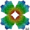
- Structure visualization
| Movie |
Movie viewer |
|---|---|
| Structure viewer | Molecule: MolmilJmol/JSmol |
- Downloads & links
-Download
| PDBx/mmCIF format | 6uea.cif.gz | 487 KB | Display | PDBx/mmCIF format |
|---|---|---|---|---|
| PDB format | pdb6uea.ent.gz | 392.9 KB | Display | PDB format |
| PDBx/mmJSON format | 6uea.json.gz | Tree view | PDBx/mmJSON format | |
| Others |  Other downloads Other downloads |
-Validation report
| Arichive directory | https://data.pdbj.org/pub/pdb/validation_reports/ue/6ueaftp://data.pdbj.org/pub/pdb/validation_reports/ue/6uea | HTTPS FTP |
|---|
-Related structure data
| Related structure data | 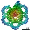 20752MC 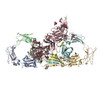 6ue7C 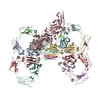 6ue8C 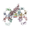 6ue9C M: map data used to model this data C: citing same article ( |
|---|---|
| Similar structure data |
-Links
 PDBj
PDBj


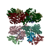
- Assembly
| Deposited unit | 
|
|---|---|
| 1 |
|
-Components
-Antibody / Protein / Immunoglobulin ... , 3 types, 12 molecules ABFGKLEHIJCD
| #1: Antibody | Mass: 26682.146 Da / Num. of mol.: 10 / Fragment: UNP residues 110-340 Source method: isolated from a genetically manipulated source Source: (gene. exp.) Homo sapiens (human) / Gene: IGHA2 / Production host:   Cricetulus griseus (Chinese hamster) / References: UniProt: P01877 Cricetulus griseus (Chinese hamster) / References: UniProt: P01877#2: Protein | | Mass: 65154.094 Da / Num. of mol.: 1 / Fragment: UNP residues 19-603 Source method: isolated from a genetically manipulated source Source: (gene. exp.) Homo sapiens (human) / Gene: PIGR / Production host: Cricetulus griseus (Chinese hamster) / References: UniProt: P01833#3: Protein | | Mass: 15611.458 Da / Num. of mol.: 1 / Fragment: UNP residues 23-159 Source method: isolated from a genetically manipulated source Source: (gene. exp.) Homo sapiens (human) / Gene: JCHAIN, IGCJ, IGJ / Production host: Cricetulus griseus (Chinese hamster) / References: UniProt: P01591 |
|---|
-Sugars , 3 types, 9 molecules 
| #4: Polysaccharide | 2-acetamido-2-deoxy-beta-D-glucopyranose-(1-4)-2-acetamido-2-deoxy-beta-D-glucopyranose Source method: isolated from a genetically manipulated source #5: Polysaccharide | Source method: isolated from a genetically manipulated source #6: Sugar |  Type: D-saccharide, beta linking / Mass: 221.208 Da / Num. of mol.: 2 Type: D-saccharide, beta linking / Mass: 221.208 Da / Num. of mol.: 2Source method: isolated from a genetically manipulated source Formula: C8H15NO6 |
|---|
-Details
| Has ligand of interest | N |
|---|---|
| Has protein modification | Y |
-Experimental details
-Experiment
| Experiment | Method: ELECTRON MICROSCOPY |
|---|---|
| EM experiment | Aggregation state: PARTICLE / 3D reconstruction method: single particle reconstruction |
- Sample preparation
| Component | Name: pentameric sIgA complex / Type: COMPLEX / Entity ID: #1-#3 / Source: RECOMBINANT |
|---|---|
| Molecular weight | Value: 0.347 MDa / Experimental value: NO |
| Source (natural) | Organism: Homo sapiens (human) |
| Source (recombinant) | Organism: Cricetulus griseus (Chinese hamster) |
| Buffer solution | pH: 7.2 |
| Specimen | Embedding applied: NO / Shadowing applied: NO / Staining applied: NO / Vitrification applied: YES |
| Specimen support | Details: unspecified |
| Vitrification | Instrument: FEI VITROBOT MARK IV / Cryogen name: ETHANE / Humidity: 100 % / Chamber temperature: 277 K |
- Electron microscopy imaging
| Experimental equipment |  Model: Titan Krios / Image courtesy: FEI Company |
|---|---|
| Microscopy | Model: FEI TITAN KRIOS |
| Electron gun | Electron source:  FIELD EMISSION GUN / Accelerating voltage: 300 kV / Illumination mode: FLOOD BEAM FIELD EMISSION GUN / Accelerating voltage: 300 kV / Illumination mode: FLOOD BEAM |
| Electron lens | Mode: BRIGHT FIELD / C2 aperture diameter: 100 µm |
| Specimen holder | Cryogen: NITROGEN / Specimen holder model: FEI TITAN KRIOS AUTOGRID HOLDER |
| Image recording | Average exposure time: 10 sec. / Electron dose: 51.91 e/Å2 / Film or detector model: GATAN K2 SUMMIT (4k x 4k) |
| EM imaging optics | Energyfilter name: GIF Bioquantum / Energyfilter slit width: 20 eV |
- Processing
| EM software |
| ||||||||||||
|---|---|---|---|---|---|---|---|---|---|---|---|---|---|
| CTF correction | Type: PHASE FLIPPING AND AMPLITUDE CORRECTION | ||||||||||||
| 3D reconstruction | Resolution: 3 Å / Resolution method: FSC 0.143 CUT-OFF / Num. of particles: 165069 / Symmetry type: POINT |
