 Movie
Movie Controller

[English] 日本語
 Yorodumi
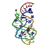
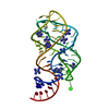
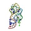
- PDB-6ubu: 1.60 A resolution structure of the guanine riboswitch bound to guanine -
+ Open data

- Basic information
| Entry | Database: PDB / ID: 6ubu | ||||||
|---|---|---|---|---|---|---|---|
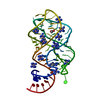
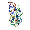
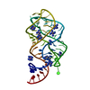
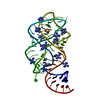
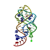
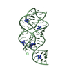
| Title | 1.60 A resolution structure of the guanine riboswitch bound to guanine | ||||||
 Components Components | Guanine riboswitch aptamer domain | ||||||
 Keywords Keywords | RNA / riboswitch aptamer guanine purine nucleobase | ||||||
| Function / homology | ACETATE ION / GUANINE / COBALT HEXAMMINE(III) / RNA / RNA (> 10) Function and homology information Function and homology information | ||||||
| Biological species |  | ||||||
| Method |  X-RAY DIFFRACTION / MOLECULAR REPLACEMENT / Resolution: 1.6 Å X-RAY DIFFRACTION / MOLECULAR REPLACEMENT / Resolution: 1.6 Å | ||||||
 Authors Authors | Matyjasik, M.M. / Batey, R.T. | ||||||
| Funding support |  United States, 1items United States, 1items
| ||||||
 Citation Citation | Journal: Molecules / Year: 2020 Title: High Affinity Binding of N2-Modified Guanine Derivatives Significantly Disrupts the Ligand Binding Pocket of the Guanine Riboswitch. Authors: Matyjasik, M.M. / Hall, S.D. / Batey, R.T. | ||||||
| History |
|
- Structure visualization
| Structure viewer | Molecule: MolmilJmol/JSmol |
|---|
- Downloads & links
-Download
| PDBx/mmCIF format | 6ubu.cif.gz | 60.4 KB | Display | PDBx/mmCIF format |
|---|---|---|---|---|
| PDB format | pdb6ubu.ent.gz | 37.9 KB | Display | PDB format |
| PDBx/mmJSON format | 6ubu.json.gz | Tree view | PDBx/mmJSON format | |
| Others |  Other downloads Other downloads |
-Validation report
| Arichive directory | https://data.pdbj.org/pub/pdb/validation_reports/ub/6ubuftp://data.pdbj.org/pub/pdb/validation_reports/ub/6ubu | HTTPS FTP |
|---|
-Related structure data
| Related structure data |  6uc7C 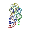 6uc8C  6uc9C  4fe5S S: Starting model for refinement C: citing same article ( |
|---|---|
| Similar structure data |


-Links
 PDBj
PDBj


- Assembly
| Deposited unit | 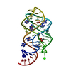
| ||||||||||
|---|---|---|---|---|---|---|---|---|---|---|---|
| 1 |
| ||||||||||
| Unit cell |
|
-Components
| #1: RNA chain | Mass: 21506.783 Da / Num. of mol.: 1 / Source method: obtained synthetically / Source: (synth.) | ||||||||
|---|---|---|---|---|---|---|---|---|---|
| #2: Chemical | ChemComp-NCO /   Mass: 161.116 Da / Num. of mol.: 7 / Source method: obtained synthetically / Formula: CoH18N6 Mass: 161.116 Da / Num. of mol.: 7 / Source method: obtained synthetically / Formula: CoH18N6#3: Chemical |   Mass: 59.044 Da / Num. of mol.: 3 / Source method: obtained synthetically / Formula: C2H3O2 Mass: 59.044 Da / Num. of mol.: 3 / Source method: obtained synthetically / Formula: C2H3O2#4: Chemical | ChemComp-GUN / |   Mass: 151.126 Da / Num. of mol.: 1 / Source method: obtained synthetically / Formula: C5H5N5O / Feature type: SUBJECT OF INVESTIGATION Mass: 151.126 Da / Num. of mol.: 1 / Source method: obtained synthetically / Formula: C5H5N5O / Feature type: SUBJECT OF INVESTIGATION#5: Water | ChemComp-HOH / |  Mass: 18.015 Da / Num. of mol.: 240 / Source method: isolated from a natural source / Formula: H2O Mass: 18.015 Da / Num. of mol.: 240 / Source method: isolated from a natural source / Formula: H2OHas ligand of interest | Y | |
-Experimental details
-Experiment
| Experiment | Method: X-RAY DIFFRACTION / Number of used crystals: 1 |
|---|
- Sample preparation
| Crystal | Density Matthews: 2.29 Å3/Da / Density % sol: 46.29 % |
|---|---|
| Crystal grow | Temperature: 297 K / Method: vapor diffusion, hanging drop / pH: 7.5 Details: 10 mM potassium HEPES pH 7.5, 20% v/v PEG 3000 MW, 40-50 mM cobalt hexamine, 600-650 mM ammonium acetate |
-Data collection
| Diffraction | Mean temperature: 100 K / Ambient temp details: Liquid nitrogen cryostream / Serial crystal experiment: N |
|---|---|
| Diffraction source | Source: ROTATING ANODE / Type: RIGAKU MICROMAX-007 HF / Wavelength: 1.5406 Å |
| Detector | Type: DECTRIS PILATUS 200K / Detector: PIXEL / Date: Apr 25, 2019 |
| Radiation | Monochromator: Cu / Protocol: SINGLE WAVELENGTH / Monochromatic (M) / Laue (L): M / Scattering type: x-ray |
| Radiation wavelength | Wavelength: 1.5406 Å / Relative weight: 1 |
| Reflection | Resolution: 1.597→19.66 Å / Num. obs: 37435 / % possible obs: 83.41 % / Redundancy: 4.5 % / Biso Wilson estimate: 20.23 Å2 / Rpim(I) all: 0.061 / Rrim(I) all: 0.131 / Rsym value: 0.131 / Χ2: 2.015 / Net I/σ(I): 13.67 |
| Reflection shell | Resolution: 1.597→1.654 Å / Redundancy: 1 % / Rmerge(I) obs: 0.361 / Mean I/σ(I) obs: 1.876 / Num. unique obs: 593 / CC1/2: 0.622 / Χ2: 2.382 / % possible all: 23.15 |
- Processing
| Software |
| ||||||||||||||||||||||||
|---|---|---|---|---|---|---|---|---|---|---|---|---|---|---|---|---|---|---|---|---|---|---|---|---|---|
| Refinement | Method to determine structure: MOLECULAR REPLACEMENT Starting model: 4FE5 Resolution: 1.6→19.66 Å / SU ML: 0.16 / Cross valid method: FREE R-VALUE / σ(F): 1.34 / Phase error: 27.37
| ||||||||||||||||||||||||
| Solvent computation | Shrinkage radii: 0.9 Å / VDW probe radii: 1.11 Å | ||||||||||||||||||||||||
| Displacement parameters | Biso mean: 28.48 Å2 | ||||||||||||||||||||||||
| Refinement step | Cycle: final / Resolution: 1.6→19.66 Å
| ||||||||||||||||||||||||
| Refine LS restraints |
|
