 Movie
Movie Controller

[English] 日本語
 Yorodumi
- PDB-3rkf: Crystal structure of guanine riboswitch C61U/G37A double mutant b... -
+ Open data

- Basic information
| Entry | Database: PDB / ID: 3rkf | ||||||
|---|---|---|---|---|---|---|---|
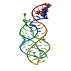
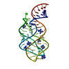
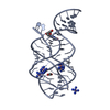
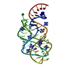
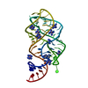
| Title | Crystal structure of guanine riboswitch C61U/G37A double mutant bound to thio-guanine | ||||||
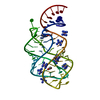
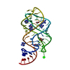
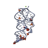
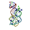
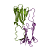
 Components Components | Guanine riboswitch | ||||||
 Keywords Keywords | RNA / three-way junction / riboswitch / m-RNA / thioguanine | ||||||
| Function / homology | 2-amino-1,9-dihydro-6H-purine-6-thione / COBALT HEXAMMINE(III) / RNA / RNA (> 10) Function and homology information Function and homology information | ||||||
| Method |  X-RAY DIFFRACTION / SYNCHROTRON / MOLECULAR REPLACEMENT / Resolution: 2.5 Å X-RAY DIFFRACTION / SYNCHROTRON / MOLECULAR REPLACEMENT / Resolution: 2.5 Å | ||||||
 Authors Authors | Buck, J. / Wacker, A. / Warkentin, E. / Woehnert, J. / Wirmer-Bartoschek, J. / Schwalbe, H. | ||||||
 Citation Citation | Journal: Nucleic Acids Res. / Year: 2011 Title: Influence of ground-state structure and Mg2+ binding on folding kinetics of the guanine-sensing riboswitch aptamer domain. Authors: Buck, J. / Wacker, A. / Warkentin, E. / Wohnert, J. / Wirmer-Bartoschek, J. / Schwalbe, H. | ||||||
| History |
|
- Structure visualization
| Structure viewer | Molecule: MolmilJmol/JSmol |
|---|
- Downloads & links
-Download
| PDBx/mmCIF format | 3rkf.cif.gz | 159 KB | Display | PDBx/mmCIF format |
|---|---|---|---|---|
| PDB format | pdb3rkf.ent.gz | 121.7 KB | Display | PDB format |
| PDBx/mmJSON format | 3rkf.json.gz | Tree view | PDBx/mmJSON format | |
| Others |  Other downloads Other downloads |
-Validation report
| Arichive directory | https://data.pdbj.org/pub/pdb/validation_reports/rk/3rkfftp://data.pdbj.org/pub/pdb/validation_reports/rk/3rkf | HTTPS FTP |
|---|
-Related structure data
| Related structure data |  1u8d S: Starting model for refinement |
|---|---|
| Similar structure data |
-Links
 PDBj
PDBj


- Assembly
| Deposited unit | 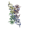
| ||||||||
|---|---|---|---|---|---|---|---|---|---|
| 1 | 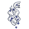
| ||||||||
| 2 | 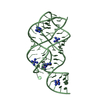
| ||||||||
| 3 | 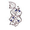
| ||||||||
| 4 | 
| ||||||||
| Unit cell |
|
-Components
| #1: RNA chain | Mass: 21523.766 Da / Num. of mol.: 4 / Mutation: C61U G37A / Source method: obtained synthetically #2: Chemical | ChemComp-DX4 / 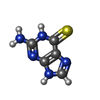  Mass: 167.192 Da / Num. of mol.: 4 / Source method: obtained synthetically / Formula: C5H5N5S / Comment: medication*YM Mass: 167.192 Da / Num. of mol.: 4 / Source method: obtained synthetically / Formula: C5H5N5S / Comment: medication*YM#3: Chemical | ChemComp-NCO /   Mass: 161.116 Da / Num. of mol.: 26 / Source method: obtained synthetically / Formula: CoH18N6 Mass: 161.116 Da / Num. of mol.: 26 / Source method: obtained synthetically / Formula: CoH18N6#4: Water | ChemComp-HOH / |  Mass: 18.015 Da / Num. of mol.: 12 / Source method: isolated from a natural source / Formula: H2O Mass: 18.015 Da / Num. of mol.: 12 / Source method: isolated from a natural source / Formula: H2O |
|---|
-Experimental details
-Experiment
| Experiment | Method: X-RAY DIFFRACTION / Number of used crystals: 1 |
|---|
- Sample preparation
| Crystal | Density Matthews: 2.42 Å3/Da / Density % sol: 49.08 % |
|---|---|
| Crystal grow | Temperature: 298 K / Method: vapor diffusion, hanging drop / pH: 7.5 Details: 5mM K+ - HEPES, 12mM [Co(NH3)6]3+, 25% polyethylene glycol 4000, 450mM ammonium acetate, pH 7.5, VAPOR DIFFUSION, HANGING DROP, temperature 298K |
-Data collection
| Diffraction | Mean temperature: 100 K |
|---|---|
| Diffraction source | Source: SYNCHROTRON / Site: SLS  / Beamline: X10SA / Wavelength: 1.02 Å / Beamline: X10SA / Wavelength: 1.02 Å |
| Detector | Type: MARMOSAIC 225 mm CCD / Detector: CCD / Date: Jun 8, 2009 |
| Radiation | Monochromator: SI 111 / Protocol: SINGLE WAVELENGTH / Monochromatic (M) / Laue (L): M / Scattering type: x-ray |
| Radiation wavelength | Wavelength: 1.02 Å / Relative weight: 1 |
| Reflection | Resolution: 2.5→44.64 Å / Num. all: 67756 / Num. obs: 26059 / % possible obs: 93.5 % / Observed criterion σ(I): -3 |
| Reflection shell | Resolution: 2.5→2.59 Å / % possible all: 82.4 |
- Processing
| Software |
| |||||||||||||||||||||||||||||||||||||||||||||||||||||||||||||||
|---|---|---|---|---|---|---|---|---|---|---|---|---|---|---|---|---|---|---|---|---|---|---|---|---|---|---|---|---|---|---|---|---|---|---|---|---|---|---|---|---|---|---|---|---|---|---|---|---|---|---|---|---|---|---|---|---|---|---|---|---|---|---|---|---|
| Refinement | Method to determine structure: MOLECULAR REPLACEMENT Starting model: PDB entry 1u8d 1u8d Resolution: 2.5→44.638 Å / σ(F): 1.96 / Phase error: 30.98 / Stereochemistry target values: TWIN_LSQ_F
| |||||||||||||||||||||||||||||||||||||||||||||||||||||||||||||||
| Solvent computation | Shrinkage radii: 0.61 Å / VDW probe radii: 0.9 Å / Solvent model: FLAT BULK SOLVENT MODEL / Bsol: 24.729 Å2 / ksol: 0.315 e/Å3 | |||||||||||||||||||||||||||||||||||||||||||||||||||||||||||||||
| Displacement parameters |
| |||||||||||||||||||||||||||||||||||||||||||||||||||||||||||||||
| Refinement step | Cycle: LAST / Resolution: 2.5→44.638 Å
| |||||||||||||||||||||||||||||||||||||||||||||||||||||||||||||||
| Refine LS restraints |
| |||||||||||||||||||||||||||||||||||||||||||||||||||||||||||||||
| LS refinement shell |
|
