 Movie
Movie Controller


+ Open data

- Basic information
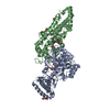
| Entry | Database: PDB / ID: 6u7a | ||||||
|---|---|---|---|---|---|---|---|
| Title | Rv3722c in complex with kynurenine | ||||||
 Components Components | Aminotransferase | ||||||
 Keywords Keywords | TRANSFERASE / Aminotransferase / Mycobacterium tuberculosis | ||||||
| Function / homology |  Function and homology information Function and homology informationaspartate transaminase / L-aspartate:2-oxoglutarate transaminase activity / extracellular region Similarity search - Function | ||||||
| Biological species |   Mycobacterium tuberculosis (bacteria) Mycobacterium tuberculosis (bacteria) | ||||||
| Method |  X-RAY DIFFRACTION / SYNCHROTRON / MOLECULAR REPLACEMENT / Resolution: 2.22 Å X-RAY DIFFRACTION / SYNCHROTRON / MOLECULAR REPLACEMENT / Resolution: 2.22 Å | ||||||
 Authors Authors | Mandyoli, L. / Sacchettini, J. | ||||||
 Citation Citation | Journal: Nat Commun / Year: 2020 Title: Aspartate aminotransferase Rv3722c governs aspartate-dependent nitrogen metabolism in Mycobacterium tuberculosis. Authors: Jansen, R.S. / Mandyoli, L. / Hughes, R. / Wakabayashi, S. / Pinkham, J.T. / Selbach, B. / Guinn, K.M. / Rubin, E.J. / Sacchettini, J.C. / Rhee, K.Y. | ||||||
| History |
|
- Structure visualization
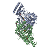
| Structure viewer | Molecule: MolmilJmol/JSmol |
|---|
- Downloads & links
-Download
| PDBx/mmCIF format | 6u7a.cif.gz | 724.1 KB | Display | PDBx/mmCIF format |
|---|---|---|---|---|
| PDB format | pdb6u7a.ent.gz | 546.9 KB | Display | PDB format |
| PDBx/mmJSON format | 6u7a.json.gz | Tree view | PDBx/mmJSON format | |
| Others |  Other downloads Other downloads |
-Validation report
| Arichive directory | https://data.pdbj.org/pub/pdb/validation_reports/u7/6u7aftp://data.pdbj.org/pub/pdb/validation_reports/u7/6u7a | HTTPS FTP |
|---|
-Related structure data
| Related structure data |  6u78C  5c6uS S: Starting model for refinement C: citing same article ( |
|---|---|
| Similar structure data |


-Links
 PDBj
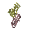
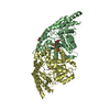
PDBj- Assembly
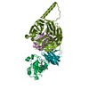
| Deposited unit | 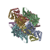
| ||||||||||||
|---|---|---|---|---|---|---|---|---|---|---|---|---|---|
| 1 | 
| ||||||||||||
| 2 | 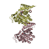
| ||||||||||||
| 3 | 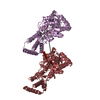
| ||||||||||||
| 4 | 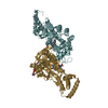
| ||||||||||||
| Unit cell |
|
-Components
-Protein , 1 types, 8 molecules ABCDEFGH
| #1: Protein | Mass: 47356.754 Da / Num. of mol.: 8 Source method: isolated from a genetically manipulated source Source: (gene. exp.) Mycobacterium tuberculosis (bacteria)Gene: DKC2_3956, DSI35_31455, ERS007661_01687, ERS007663_01945, ERS007665_01586, ERS007670_01863, ERS007672_01473, ERS007679_00092, ERS007681_00098, ERS007688_00789, ERS007703_00081, ERS007720_02793, ...Gene: DKC2_3956, DSI35_31455, ERS007661_01687, ERS007663_01945, ERS007665_01586, ERS007670_01863, ERS007672_01473, ERS007679_00092, ERS007681_00098, ERS007688_00789, ERS007703_00081, ERS007720_02793, ERS007722_01790, ERS023446_00155, ERS024276_02601, ERS027646_02441, ERS027652_01615, ERS027653_01450, ERS027656_00154, ERS027666_02093, ERS031537_01226, ERS124361_00578, EUB07_12880, EUB11_07270, EUB16_18395, SAMEA2682835_06491, SAMEA2682864_02982, SAMEA2683035_02356 Production host: |
|---|
-Non-polymers , 8 types, 1079 molecules 


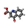


| #2: Chemical | ChemComp-Q0P / ( Mass: 437.340 Da / Num. of mol.: 1 / Source method: obtained synthetically / Formula: C18H20N3O8P / Feature type: SUBJECT OF INVESTIGATION Mass: 437.340 Da / Num. of mol.: 1 / Source method: obtained synthetically / Formula: C18H20N3O8P / Feature type: SUBJECT OF INVESTIGATION | ||||||||||||
|---|---|---|---|---|---|---|---|---|---|---|---|---|---|
| #3: Chemical | ChemComp-PO4 /  Mass: 94.971 Da / Num. of mol.: 6 / Source method: obtained synthetically / Formula: PO4 Mass: 94.971 Da / Num. of mol.: 6 / Source method: obtained synthetically / Formula: PO4#4: Chemical | ChemComp-GOL /  Mass: 92.094 Da / Num. of mol.: 10 / Source method: obtained synthetically / Formula: C3H8O3 Mass: 92.094 Da / Num. of mol.: 10 / Source method: obtained synthetically / Formula: C3H8O3#5: Chemical | ChemComp-1PE /  Mass: 238.278 Da / Num. of mol.: 6 / Source method: isolated from a natural source / Formula: C10H22O6 / Comment: precipitant*YM Mass: 238.278 Da / Num. of mol.: 6 / Source method: isolated from a natural source / Formula: C10H22O6 / Comment: precipitant*YM#6: Chemical | ChemComp-KYA /  Mass: 189.167 Da / Num. of mol.: 4 / Source method: obtained synthetically / Formula: C10H7NO3 / Feature type: SUBJECT OF INVESTIGATION Mass: 189.167 Da / Num. of mol.: 4 / Source method: obtained synthetically / Formula: C10H7NO3 / Feature type: SUBJECT OF INVESTIGATION#7: Chemical | ChemComp-PMP /  Mass: 248.173 Da / Num. of mol.: 4 / Source method: obtained synthetically / Formula: C8H13N2O5P Mass: 248.173 Da / Num. of mol.: 4 / Source method: obtained synthetically / Formula: C8H13N2O5P#8: Chemical |  Mass: 247.142 Da / Num. of mol.: 3 / Source method: obtained synthetically / Formula: C8H10NO6P Mass: 247.142 Da / Num. of mol.: 3 / Source method: obtained synthetically / Formula: C8H10NO6P#9: Water | ChemComp-HOH / | Mass: 18.015 Da / Num. of mol.: 1045 / Source method: isolated from a natural source / Formula: H2O |
-Details
| Has ligand of interest | Y |
|---|
-Experimental details
-Experiment
| Experiment | Method: X-RAY DIFFRACTION / Number of used crystals: 1 |
|---|
- Sample preparation
| Crystal | Density Matthews: 2.94 Å3/Da / Density % sol: 58.18 % |
|---|---|
| Crystal grow | Temperature: 290 K / Method: vapor diffusion, sitting drop / pH: 4.2 Details: 100 mM sodium phosphate dibasic / citric acid, 40% ethanol, 5% PEG1000 |
-Data collection
| Diffraction | Mean temperature: 100 K / Serial crystal experiment: N |
|---|---|
| Diffraction source | Source: SYNCHROTRON / Site: APS  / Beamline: 23-ID-D / Wavelength: 0.978 Å / Beamline: 23-ID-D / Wavelength: 0.978 Å |
| Detector | Type: DECTRIS PILATUS3 S 6M / Detector: PIXEL / Date: Jul 2, 2019 |
| Radiation | Monochromator: double crystal Si(111) / Protocol: SINGLE WAVELENGTH / Monochromatic (M) / Laue (L): M / Scattering type: x-ray |
| Radiation wavelength | Wavelength: 0.978 Å / Relative weight: 1 |
| Reflection | Resolution: 2.22→48.361 Å / Num. obs: 178462 / % possible obs: 89.5 % / Redundancy: 7.5 % / Biso Wilson estimate: 30.89 Å2 / CC1/2: 0.996 / Rmerge(I) obs: 0.138 / Net I/σ(I): 10.7 |
| Reflection shell | Resolution: 2.223→2.228 Å / Num. unique obs: 7803 / CC1/2: 0.63 |
- Processing
| Software |
| |||||||||||||||||||||||||||||||||||||||||||||||||||||||||||||||||||||||||||||||||||||||||||||||||||||||||||||||||||||||||||||||||||||||||||||||||||||||||||||||||||||||||||||||||||||||||||||||||||||||||||||||||||||||||
|---|---|---|---|---|---|---|---|---|---|---|---|---|---|---|---|---|---|---|---|---|---|---|---|---|---|---|---|---|---|---|---|---|---|---|---|---|---|---|---|---|---|---|---|---|---|---|---|---|---|---|---|---|---|---|---|---|---|---|---|---|---|---|---|---|---|---|---|---|---|---|---|---|---|---|---|---|---|---|---|---|---|---|---|---|---|---|---|---|---|---|---|---|---|---|---|---|---|---|---|---|---|---|---|---|---|---|---|---|---|---|---|---|---|---|---|---|---|---|---|---|---|---|---|---|---|---|---|---|---|---|---|---|---|---|---|---|---|---|---|---|---|---|---|---|---|---|---|---|---|---|---|---|---|---|---|---|---|---|---|---|---|---|---|---|---|---|---|---|---|---|---|---|---|---|---|---|---|---|---|---|---|---|---|---|---|---|---|---|---|---|---|---|---|---|---|---|---|---|---|---|---|---|---|---|---|---|---|---|---|---|---|---|---|---|---|---|---|---|
| Refinement | Method to determine structure: MOLECULAR REPLACEMENT Starting model: PDB entry 5C6U Resolution: 2.22→32.46 Å / SU ML: 0.2683 / Cross valid method: FREE R-VALUE / σ(F): 1.95 / Phase error: 24.5626 Stereochemistry target values: GeoStd + Monomer Library + CDL v1.2
| |||||||||||||||||||||||||||||||||||||||||||||||||||||||||||||||||||||||||||||||||||||||||||||||||||||||||||||||||||||||||||||||||||||||||||||||||||||||||||||||||||||||||||||||||||||||||||||||||||||||||||||||||||||||||
| Solvent computation | Shrinkage radii: 0.9 Å / VDW probe radii: 1.11 Å / Solvent model: FLAT BULK SOLVENT MODEL | |||||||||||||||||||||||||||||||||||||||||||||||||||||||||||||||||||||||||||||||||||||||||||||||||||||||||||||||||||||||||||||||||||||||||||||||||||||||||||||||||||||||||||||||||||||||||||||||||||||||||||||||||||||||||
| Displacement parameters | Biso mean: 36.9 Å2 | |||||||||||||||||||||||||||||||||||||||||||||||||||||||||||||||||||||||||||||||||||||||||||||||||||||||||||||||||||||||||||||||||||||||||||||||||||||||||||||||||||||||||||||||||||||||||||||||||||||||||||||||||||||||||
| Refinement step | Cycle: LAST / Resolution: 2.22→32.46 Å
| |||||||||||||||||||||||||||||||||||||||||||||||||||||||||||||||||||||||||||||||||||||||||||||||||||||||||||||||||||||||||||||||||||||||||||||||||||||||||||||||||||||||||||||||||||||||||||||||||||||||||||||||||||||||||
| Refine LS restraints |
| |||||||||||||||||||||||||||||||||||||||||||||||||||||||||||||||||||||||||||||||||||||||||||||||||||||||||||||||||||||||||||||||||||||||||||||||||||||||||||||||||||||||||||||||||||||||||||||||||||||||||||||||||||||||||
| LS refinement shell |
|
