 Movie
Movie Controller


+ Open data

- Basic information
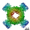
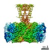
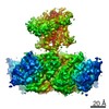
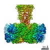
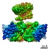
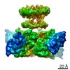
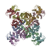
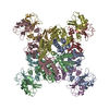
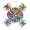
| Entry | Database: PDB / ID: 6u6h | ||||||
|---|---|---|---|---|---|---|---|
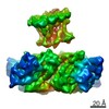
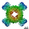
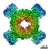
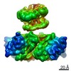
| Title | Calcium-bound MthK open-inactivated state 3 | ||||||
 Components Components | Calcium-gated potassium channel MthK | ||||||
 Keywords Keywords | TRANSPORT PROTEIN / MthK | ||||||
| Function / homology |  Function and homology information Function and homology informationmonoatomic cation transmembrane transporter activity / potassium ion transport / metal ion binding / identical protein binding / plasma membrane Similarity search - Function | ||||||
| Biological species |   Methanothermobacter thermautotrophicus (archaea) Methanothermobacter thermautotrophicus (archaea) | ||||||
| Method | ELECTRON MICROSCOPY / single particle reconstruction / cryo EM / Resolution: 5 Å | ||||||
 Authors Authors | Chen, F. / Crina, N. | ||||||
 Citation Citation | Journal: Nature / Year: 2020 Title: Ball-and-chain inactivation in a calcium-gated potassium channel. Authors: Chen Fan / Nattakan Sukomon / Emelie Flood / Jan Rheinberger / Toby W Allen / Crina M Nimigean /    Abstract: Inactivation is the process by which ion channels terminate ion flux through their pores while the opening stimulus is still present. In neurons, inactivation of both sodium and potassium channels is ...Inactivation is the process by which ion channels terminate ion flux through their pores while the opening stimulus is still present. In neurons, inactivation of both sodium and potassium channels is crucial for the generation of action potentials and regulation of firing frequency. A cytoplasmic domain of either the channel or an accessory subunit is thought to plug the open pore to inactivate the channel via a 'ball-and-chain' mechanism. Here we use cryo-electron microscopy to identify the molecular gating mechanism in calcium-activated potassium channels by obtaining structures of the MthK channel from Methanobacterium thermoautotrophicum-a purely calcium-gated and inactivating channel-in a lipid environment. In the absence of Ca, we obtained a single structure in a closed state, which was shown by atomistic simulations to be highly flexible in lipid bilayers at ambient temperature, with large rocking motions of the gating ring and bending of pore-lining helices. In Ca-bound conditions, we obtained several structures, including multiple open-inactivated conformations, further indication of a highly dynamic protein. These different channel conformations are distinguished by rocking of the gating rings with respect to the transmembrane region, indicating symmetry breakage across the channel. Furthermore, in all conformations displaying open channel pores, the N terminus of one subunit of the channel tetramer sticks into the pore and plugs it, with free energy simulations showing that this is a strong interaction. Deletion of this N terminus leads to functionally non-inactivating channels and structures of open states without a pore plug, indicating that this previously unresolved N-terminal peptide is responsible for a ball-and-chain inactivation mechanism. | ||||||
| History |
|
- Structure visualization
| Movie |
Movie viewer |
|---|---|
| Structure viewer | Molecule: MolmilJmol/JSmol |
- Downloads & links
-Download
| PDBx/mmCIF format | 6u6h.cif.gz | 290.5 KB | Display | PDBx/mmCIF format |
|---|---|---|---|---|
| PDB format | pdb6u6h.ent.gz | 203.9 KB | Display | PDB format |
| PDBx/mmJSON format | 6u6h.json.gz | Tree view | PDBx/mmJSON format | |
| Others |  Other downloads Other downloads |
-Validation report
| Arichive directory | https://data.pdbj.org/pub/pdb/validation_reports/u6/6u6hftp://data.pdbj.org/pub/pdb/validation_reports/u6/6u6h | HTTPS FTP |
|---|
-Related structure data
| Related structure data | 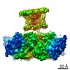 20665MC 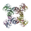 6u5nC 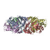 6u5pC 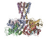 6u5rC 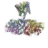 6u68C 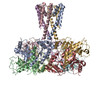 6u6dC 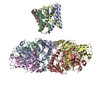 6u6eC 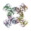 6uwnC 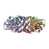 6ux4C 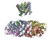 6ux7C 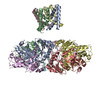 6uxaC 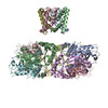 6uxbC M: map data used to model this data C: citing same article ( |
|---|---|
| Similar structure data |


-Links
 PDBj
PDBj

- Assembly
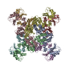
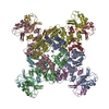
| Deposited unit | 
|
|---|---|
| 1 |
|
-Components
| #1: Protein | Mass: 37356.160 Da / Num. of mol.: 8 Source method: isolated from a genetically manipulated source Source: (gene. exp.) Methanothermobacter thermautotrophicus (archaea)Gene: mthK, MTH_1520 / Production host:  |
|---|
-Experimental details
-Experiment
| Experiment | Method: ELECTRON MICROSCOPY |
|---|---|
| EM experiment | Aggregation state: PARTICLE / 3D reconstruction method: single particle reconstruction |
- Sample preparation
| Component | Name: Calcium-bound MthK / Type: COMPLEX / Entity ID: all / Source: RECOMBINANT |
|---|---|
| Source (natural) | Organism: Methanothermobacter thermautotrophicus (archaea) |
| Source (recombinant) | Organism: |
| Buffer solution | pH: 8.5 |
| Specimen | Embedding applied: NO / Shadowing applied: NO / Staining applied: NO / Vitrification applied: YES |
| Specimen support | Details: unspecified |
| Vitrification | Cryogen name: ETHANE |
- Electron microscopy imaging
| Experimental equipment |  Model: Titan Krios / Image courtesy: FEI Company |
|---|---|
| Microscopy | Model: FEI TITAN KRIOS |
| Electron gun | Electron source:  FIELD EMISSION GUN / Accelerating voltage: 300 kV / Illumination mode: FLOOD BEAM FIELD EMISSION GUN / Accelerating voltage: 300 kV / Illumination mode: FLOOD BEAM |
| Electron lens | Mode: BRIGHT FIELD |
| Image recording | Average exposure time: 8 sec. / Electron dose: 52 e/Å2 / Film or detector model: GATAN K2 SUMMIT (4k x 4k) |
- Processing
| EM software |
| ||||||||||||||||||||||||||||||||
|---|---|---|---|---|---|---|---|---|---|---|---|---|---|---|---|---|---|---|---|---|---|---|---|---|---|---|---|---|---|---|---|---|---|
| CTF correction | Type: PHASE FLIPPING ONLY | ||||||||||||||||||||||||||||||||
| Symmetry | Point symmetry: C1 (asymmetric) | ||||||||||||||||||||||||||||||||
| 3D reconstruction | Resolution: 5 Å / Resolution method: FSC 0.143 CUT-OFF / Num. of particles: 15144 / Symmetry type: POINT |
