 Movie
Movie Controller

[English] 日本語
 Yorodumi
- PDB-6u22: Crystal structure of SFTI-triazole inhibitor in complex with beta... -
+ Open data

- Basic information
| Entry | Database: PDB / ID: 6u22 | ||||||
|---|---|---|---|---|---|---|---|
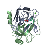
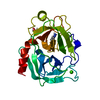
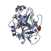
| Title | Crystal structure of SFTI-triazole inhibitor in complex with beta-trypsin | ||||||
 Components Components |
| ||||||
 Keywords Keywords | HYDROLASE/INHIBITOR / Triazole / Peptidomimetic / BIOSYNTHETIC PROTEIN / HYDROLASE-INHIBITOR complex | ||||||
| Function / homology |  Function and homology information Function and homology informationendopeptidase inhibitor activity / trypsin / serpin family protein binding / serine protease inhibitor complex / digestion / serine-type endopeptidase inhibitor activity / protease binding / endopeptidase activity / serine-type endopeptidase activity / proteolysis ...endopeptidase inhibitor activity / trypsin / serpin family protein binding / serine protease inhibitor complex / digestion / serine-type endopeptidase inhibitor activity / protease binding / endopeptidase activity / serine-type endopeptidase activity / proteolysis / : / metal ion binding Similarity search - Function | ||||||
| Biological species |   | ||||||
| Method |  X-RAY DIFFRACTION / SYNCHROTRON / MOLECULAR REPLACEMENT / Resolution: 1.42 Å X-RAY DIFFRACTION / SYNCHROTRON / MOLECULAR REPLACEMENT / Resolution: 1.42 Å | ||||||
 Authors Authors | White, A.M. / King, G.J. / Durek, T. / Craik, D.J. | ||||||
| Funding support |  Australia, 1items Australia, 1items
| ||||||
 Citation Citation | Journal: Angew.Chem.Int.Ed.Engl. / Year: 2020 Title: Application and Structural Analysis of Triazole-Bridged Disulfide Mimetics in Cyclic Peptides. Authors: White, A.M. / de Veer, S.J. / Wu, G. / Harvey, P.J. / Yap, K. / King, G.J. / Swedberg, J.E. / Wang, C.K. / Law, R.H.P. / Durek, T. / Craik, D.J. | ||||||
| History |
|
- Structure visualization
| Structure viewer | Molecule: MolmilJmol/JSmol |
|---|
- Downloads & links
-Download
| PDBx/mmCIF format | 6u22.cif.gz | 142.3 KB | Display | PDBx/mmCIF format |
|---|---|---|---|---|
| PDB format | pdb6u22.ent.gz | 110.2 KB | Display | PDB format |
| PDBx/mmJSON format | 6u22.json.gz | Tree view | PDBx/mmJSON format | |
| Others |  Other downloads Other downloads |
-Validation report
| Arichive directory | https://data.pdbj.org/pub/pdb/validation_reports/u2/6u22ftp://data.pdbj.org/pub/pdb/validation_reports/u2/6u22 | HTTPS FTP |
|---|
-Related structure data
| Related structure data | 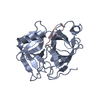 6q1uC  6u24C  6u7qC  6u7rC  6u7sC  6u7uC  6u7wC  6u7xC  6vxyC  6vy8C  1sfiS S: Starting model for refinement C: citing same article ( |
|---|---|
| Similar structure data |


-Links
 PDBj
PDBj

- Assembly
| Deposited unit | 
| ||||||||
|---|---|---|---|---|---|---|---|---|---|
| 1 |
| ||||||||
| Unit cell |
|
-Components
| #1: Protein | Mass: 23324.287 Da / Num. of mol.: 1 / Source method: isolated from a natural source / Source: (natural) |
|---|---|
| #2: Protein/peptide | Mass: 1471.699 Da / Num. of mol.: 1 / Source method: obtained synthetically / Source: (synth.) |
| #3: Chemical | ChemComp-CA /   Mass: 40.078 Da / Num. of mol.: 1 / Source method: obtained synthetically / Formula: Ca Mass: 40.078 Da / Num. of mol.: 1 / Source method: obtained synthetically / Formula: Ca |
| #4: Chemical | ChemComp-WMH /   Mass: 83.092 Da / Num. of mol.: 1 / Source method: obtained synthetically / Formula: C3H5N3 / Feature type: SUBJECT OF INVESTIGATION Mass: 83.092 Da / Num. of mol.: 1 / Source method: obtained synthetically / Formula: C3H5N3 / Feature type: SUBJECT OF INVESTIGATION |
| #5: Water | ChemComp-HOH /  Mass: 18.015 Da / Num. of mol.: 198 / Source method: isolated from a natural source / Formula: H2O Mass: 18.015 Da / Num. of mol.: 198 / Source method: isolated from a natural source / Formula: H2O |
| Has ligand of interest | Y |
| Has protein modification | Y |
-Experimental details
-Experiment
| Experiment | Method: X-RAY DIFFRACTION / Number of used crystals: 1 |
|---|
- Sample preparation
| Crystal | Density Matthews: 2.77 Å3/Da / Density % sol: 55.64 % |
|---|---|
| Crystal grow | Temperature: 277 K / Method: vapor diffusion, hanging drop / pH: 8.5 Details: Precipitant: PEG4000 (25%-35% w/v), TrisBase (0.1 M, 8.5-9.5 pH) and NH4Ac (0.26 M). Protein solution: 22 mg/mL in Tris.HCl (0.1 M, pH 8.0) with CaCl2 (10 mM) PH range: 8.5-9.5 |
-Data collection
| Diffraction | Mean temperature: 100 K / Serial crystal experiment: N |
|---|---|
| Diffraction source | Source: SYNCHROTRON / Site: Australian Synchrotron / Beamline: MX2 / Wavelength: 0.95373 Å |
| Detector | Type: ADSC QUANTUM 315r / Detector: CCD / Date: Jun 27, 2019 |
| Radiation | Protocol: SINGLE WAVELENGTH / Monochromatic (M) / Laue (L): M / Scattering type: x-ray |
| Radiation wavelength | Wavelength: 0.95373 Å / Relative weight: 1 |
| Reflection | Resolution: 1.42→46.85 Å / Num. obs: 52571 / % possible obs: 99.5 % / Redundancy: 13.2 % / Rmerge(I) obs: 0.068 / Net I/σ(I): 18.2 |
| Reflection shell | Resolution: 1.42→1.44 Å / Rmerge(I) obs: 0.371 / Num. unique obs: 2321 |
- Processing
| Software |
| ||||||||||||||||||||||||||||||||||||||||||||||||||||||||||||||||||||||||||||||||||||||||||
|---|---|---|---|---|---|---|---|---|---|---|---|---|---|---|---|---|---|---|---|---|---|---|---|---|---|---|---|---|---|---|---|---|---|---|---|---|---|---|---|---|---|---|---|---|---|---|---|---|---|---|---|---|---|---|---|---|---|---|---|---|---|---|---|---|---|---|---|---|---|---|---|---|---|---|---|---|---|---|---|---|---|---|---|---|---|---|---|---|---|---|---|
| Refinement | Method to determine structure: MOLECULAR REPLACEMENT Starting model: 1SFI Resolution: 1.42→37.462 Å / SU ML: 0.1 / Cross valid method: THROUGHOUT / σ(F): 1.4 / Phase error: 15.46 / Stereochemistry target values: ML
| ||||||||||||||||||||||||||||||||||||||||||||||||||||||||||||||||||||||||||||||||||||||||||
| Solvent computation | Shrinkage radii: 0.9 Å / VDW probe radii: 1.11 Å / Solvent model: FLAT BULK SOLVENT MODEL | ||||||||||||||||||||||||||||||||||||||||||||||||||||||||||||||||||||||||||||||||||||||||||
| Displacement parameters | Biso max: 60.15 Å2 / Biso mean: 21.3542 Å2 / Biso min: 10.45 Å2 | ||||||||||||||||||||||||||||||||||||||||||||||||||||||||||||||||||||||||||||||||||||||||||
| Refinement step | Cycle: final / Resolution: 1.42→37.462 Å
| ||||||||||||||||||||||||||||||||||||||||||||||||||||||||||||||||||||||||||||||||||||||||||
| LS refinement shell | Refine-ID: X-RAY DIFFRACTION / Rfactor Rfree error: 0
| ||||||||||||||||||||||||||||||||||||||||||||||||||||||||||||||||||||||||||||||||||||||||||
| Refinement TLS params. | Method: refined / Refine-ID: X-RAY DIFFRACTION
| ||||||||||||||||||||||||||||||||||||||||||||||||||||||||||||||||||||||||||||||||||||||||||
| Refinement TLS group |
|
