 Movie
Movie Controller

[English] 日本語
 Yorodumi
- PDB-6u1k: Thermus thermophilus D-alanine-D-alanine ligase in complex with A... -
+ Open data

- Basic information
| Entry | Database: PDB / ID: 6u1k | ||||||
|---|---|---|---|---|---|---|---|
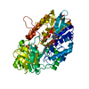
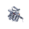
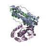
| Title | Thermus thermophilus D-alanine-D-alanine ligase in complex with ADP, carbonate, D-alanine-D-alanine, Mg2+ and K+ | ||||||
 Components Components | D-alanine--D-alanine ligase | ||||||
 Keywords Keywords | LIGASE / ATP-grasp domain / ATP binding / metal binding | ||||||
| Function / homology |  Function and homology information Function and homology informationD-alanine-D-alanine ligase / D-alanine-D-alanine ligase activity / peptidoglycan biosynthetic process / cell wall organization / regulation of cell shape / ATP binding / metal ion binding / cytosol Similarity search - Function | ||||||
| Biological species |   Thermus thermophilus (bacteria) Thermus thermophilus (bacteria) | ||||||
| Method |  X-RAY DIFFRACTION / SYNCHROTRON / MOLECULAR REPLACEMENT / molecular replacement / Resolution: 1.67 Å X-RAY DIFFRACTION / SYNCHROTRON / MOLECULAR REPLACEMENT / molecular replacement / Resolution: 1.67 Å | ||||||
 Authors Authors | Pederick, J.L. / Bruning, J.B. | ||||||
 Citation Citation | Journal: J.Biol.Chem. / Year: 2020 Title: d-Alanine-d-alanine ligase as a model for the activation of ATP-grasp enzymes by monovalent cations. Authors: Pederick, J.L. / Thompson, A.P. / Bell, S.G. / Bruning, J.B. | ||||||
| History |
|
- Structure visualization
| Structure viewer | Molecule: MolmilJmol/JSmol |
|---|
- Downloads & links
-Download
| PDBx/mmCIF format | 6u1k.cif.gz | 280.9 KB | Display | PDBx/mmCIF format |
|---|---|---|---|---|
| PDB format | pdb6u1k.ent.gz | 218.9 KB | Display | PDB format |
| PDBx/mmJSON format | 6u1k.json.gz | Tree view | PDBx/mmJSON format | |
| Others |  Other downloads Other downloads |
-Validation report
| Arichive directory | https://data.pdbj.org/pub/pdb/validation_reports/u1/6u1kftp://data.pdbj.org/pub/pdb/validation_reports/u1/6u1k | HTTPS FTP |
|---|
-Related structure data
| Related structure data | 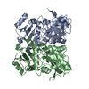 6u1cC 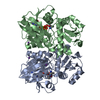 6u1dC 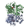 6u1eC 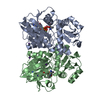 6u1fC 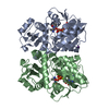 6u1gC  6u1hC 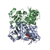 6u1iC 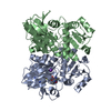 6u1jC  2zdhS C: citing same article ( S: Starting model for refinement |
|---|---|
| Similar structure data |


-Links
 PDBj
PDBj


- Assembly
| Deposited unit | 
| ||||||||
|---|---|---|---|---|---|---|---|---|---|
| 1 | 
| ||||||||
| 2 | 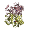
| ||||||||
| Unit cell |
|
-Components
-Protein , 1 types, 4 molecules ABCD
| #1: Protein | Mass: 35788.988 Da / Num. of mol.: 4 Source method: isolated from a genetically manipulated source Source: (gene. exp.) Thermus thermophilus (strain HB8 / ATCC 27634 / DSM 579) (bacteria)Strain: HB8 / ATCC 27634 / DSM 579 / Gene: ddl, TTHA1587 / Production host: |
|---|
-Non-polymers , 6 types, 832 molecules 


| #2: Chemical | ChemComp-DAL /  Type: D-peptide linking / Mass: 89.093 Da / Num. of mol.: 8 / Source method: obtained synthetically / Formula: C3H7NO2 Type: D-peptide linking / Mass: 89.093 Da / Num. of mol.: 8 / Source method: obtained synthetically / Formula: C3H7NO2#3: Chemical | ChemComp-MG /  Mass: 24.305 Da / Num. of mol.: 8 / Source method: obtained synthetically / Formula: Mg Mass: 24.305 Da / Num. of mol.: 8 / Source method: obtained synthetically / Formula: Mg#4: Chemical | ChemComp-K /  Mass: 39.098 Da / Num. of mol.: 4 / Source method: isolated from a natural source / Formula: K Mass: 39.098 Da / Num. of mol.: 4 / Source method: isolated from a natural source / Formula: K#5: Chemical | ChemComp-ADP /  Mass: 427.201 Da / Num. of mol.: 4 / Source method: obtained synthetically / Formula: C10H15N5O10P2 / Comment: ADP, energy-carrying molecule*YM Mass: 427.201 Da / Num. of mol.: 4 / Source method: obtained synthetically / Formula: C10H15N5O10P2 / Comment: ADP, energy-carrying molecule*YM#6: Chemical | ChemComp-CO3 /  Mass: 60.009 Da / Num. of mol.: 4 / Source method: obtained synthetically / Formula: CO3 Mass: 60.009 Da / Num. of mol.: 4 / Source method: obtained synthetically / Formula: CO3#7: Water | ChemComp-HOH / | Mass: 18.015 Da / Num. of mol.: 804 / Source method: isolated from a natural source / Formula: H2O |
|---|
-Details
| Has ligand of interest | N |
|---|
-Experimental details
-Experiment
| Experiment | Method: X-RAY DIFFRACTION / Number of used crystals: 1 |
|---|
- Sample preparation
| Crystal | Density Matthews: 2.03 Å3/Da / Density % sol: 39.55 % |
|---|---|
| Crystal grow | Temperature: 289.15 K / Method: vapor diffusion, hanging drop / pH: 6.8 Details: 15-18% PEG 3350, 100 mM Bis-Tris pH 6.8, 100 mM magnesium formate |
-Data collection
| Diffraction | Mean temperature: 100 K / Serial crystal experiment: N | ||||||||||||||||||||||||||||||
|---|---|---|---|---|---|---|---|---|---|---|---|---|---|---|---|---|---|---|---|---|---|---|---|---|---|---|---|---|---|---|---|
| Diffraction source | Source: SYNCHROTRON / Site: Australian Synchrotron  / Beamline: MX2 / Wavelength: 0.9537 Å / Beamline: MX2 / Wavelength: 0.9537 Å | ||||||||||||||||||||||||||||||
| Detector | Type: DECTRIS EIGER X 16M / Detector: PIXEL / Date: Mar 30, 2019 | ||||||||||||||||||||||||||||||
| Radiation | Protocol: SINGLE WAVELENGTH / Monochromatic (M) / Laue (L): M / Scattering type: x-ray | ||||||||||||||||||||||||||||||
| Radiation wavelength | Wavelength: 0.9537 Å / Relative weight: 1 | ||||||||||||||||||||||||||||||
| Reflection | Resolution: 1.67→48.15 Å / Num. obs: 127289 / % possible obs: 96.8 % / Redundancy: 3.4 % / CC1/2: 0.995 / Rmerge(I) obs: 0.117 / Rpim(I) all: 0.074 / Rrim(I) all: 0.139 / Net I/σ(I): 6.2 / Num. measured all: 430702 / Scaling rejects: 3 | ||||||||||||||||||||||||||||||
| Reflection shell | Diffraction-ID: 1
|
-Phasing
| Phasing | Method: molecular replacement | |||||||||
|---|---|---|---|---|---|---|---|---|---|---|
| Phasing MR |
|
- Processing
| Software |
| ||||||||||||||||||||||||||||||||||||||||||||||||||||||||||||||||||||||||||||||||||||||||||||||||||||||||||||||||||||||||||||||||||||||||||||||||||||||||||||||||||||||||||||||||||||||||||
|---|---|---|---|---|---|---|---|---|---|---|---|---|---|---|---|---|---|---|---|---|---|---|---|---|---|---|---|---|---|---|---|---|---|---|---|---|---|---|---|---|---|---|---|---|---|---|---|---|---|---|---|---|---|---|---|---|---|---|---|---|---|---|---|---|---|---|---|---|---|---|---|---|---|---|---|---|---|---|---|---|---|---|---|---|---|---|---|---|---|---|---|---|---|---|---|---|---|---|---|---|---|---|---|---|---|---|---|---|---|---|---|---|---|---|---|---|---|---|---|---|---|---|---|---|---|---|---|---|---|---|---|---|---|---|---|---|---|---|---|---|---|---|---|---|---|---|---|---|---|---|---|---|---|---|---|---|---|---|---|---|---|---|---|---|---|---|---|---|---|---|---|---|---|---|---|---|---|---|---|---|---|---|---|---|---|---|---|
| Refinement | Method to determine structure: MOLECULAR REPLACEMENT Starting model: 2ZDH Resolution: 1.67→43.575 Å / SU ML: 0.27 / Cross valid method: THROUGHOUT / σ(F): 1.96 / Phase error: 29.39 / Stereochemistry target values: ML
| ||||||||||||||||||||||||||||||||||||||||||||||||||||||||||||||||||||||||||||||||||||||||||||||||||||||||||||||||||||||||||||||||||||||||||||||||||||||||||||||||||||||||||||||||||||||||||
| Solvent computation | Shrinkage radii: 0.9 Å / VDW probe radii: 1.11 Å / Solvent model: FLAT BULK SOLVENT MODEL | ||||||||||||||||||||||||||||||||||||||||||||||||||||||||||||||||||||||||||||||||||||||||||||||||||||||||||||||||||||||||||||||||||||||||||||||||||||||||||||||||||||||||||||||||||||||||||
| Displacement parameters | Biso max: 68.64 Å2 / Biso mean: 23.9075 Å2 / Biso min: 7.97 Å2 | ||||||||||||||||||||||||||||||||||||||||||||||||||||||||||||||||||||||||||||||||||||||||||||||||||||||||||||||||||||||||||||||||||||||||||||||||||||||||||||||||||||||||||||||||||||||||||
| Refinement step | Cycle: final / Resolution: 1.67→43.575 Å
| ||||||||||||||||||||||||||||||||||||||||||||||||||||||||||||||||||||||||||||||||||||||||||||||||||||||||||||||||||||||||||||||||||||||||||||||||||||||||||||||||||||||||||||||||||||||||||
| LS refinement shell | Refine-ID: X-RAY DIFFRACTION / Rfactor Rfree error: 0
|
