 Movie
Movie Controller

[English] 日本語
 Yorodumi
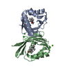
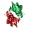
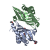
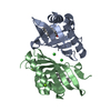
- PDB-6tzd: Crystal Structure of Ketosteroid Isomerase from Pseudomonas Putid... -
+ Open data

- Basic information
| Entry | Database: PDB / ID: 6tzd | ||||||
|---|---|---|---|---|---|---|---|
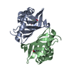
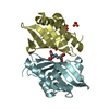
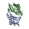
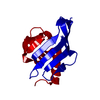
| Title | Crystal Structure of Ketosteroid Isomerase from Pseudomonas Putida (pKSI) bound to 4-Androstenedione at 280 K | ||||||
 Components Components | Steroid Delta-isomerase | ||||||
 Keywords Keywords | ISOMERASE | ||||||
| Function / homology | Ketosteroid isomerase / steroid Delta-isomerase / SnoaL-like domain / steroid Delta-isomerase activity / SnoaL-like domain / steroid metabolic process / NTF2-like domain superfamily / 4-ANDROSTENE-3-17-DIONE / Steroid Delta-isomerase Function and homology information Function and homology information | ||||||
| Biological species |  Pseudomonas putida (bacteria) Pseudomonas putida (bacteria) | ||||||
| Method |  X-RAY DIFFRACTION / SYNCHROTRON / MOLECULAR REPLACEMENT / Resolution: 1.4507 Å X-RAY DIFFRACTION / SYNCHROTRON / MOLECULAR REPLACEMENT / Resolution: 1.4507 Å | ||||||
 Authors Authors | Yabukarski, F. / Herschlag, D. | ||||||
| Funding support |  United States, 1items United States, 1items
| ||||||
 Citation Citation | Journal: Proc.Natl.Acad.Sci.USA / Year: 2020 Title: Assessment of enzyme active site positioning and tests of catalytic mechanisms through X-ray-derived conformational ensembles. Authors: Yabukarski, F. / Biel, J.T. / Pinney, M.M. / Doukov, T. / Powers, A.S. / Fraser, J.S. / Herschlag, D. | ||||||
| History |
|
- Structure visualization
| Structure viewer | Molecule: MolmilJmol/JSmol |
|---|
- Downloads & links
-Download
| PDBx/mmCIF format | 6tzd.cif.gz | 180.1 KB | Display | PDBx/mmCIF format |
|---|---|---|---|---|
| PDB format | pdb6tzd.ent.gz | 146.1 KB | Display | PDB format |
| PDBx/mmJSON format | 6tzd.json.gz | Tree view | PDBx/mmJSON format | |
| Others |  Other downloads Other downloads |
-Validation report
| Summary document | 6tzd_validation.pdf.gz | 367.8 KB | Display | wwPDB validaton report |
|---|---|---|---|---|
| Full document | 6tzd_full_validation.pdf.gz | 364.8 KB | Display | |
| Data in XML | 6tzd_validation.xml.gz | 1.4 KB | Display | |
| Data in CIF | 6tzd_validation.cif.gz | 4.9 KB | Display | |
| Arichive directory | https://data.pdbj.org/pub/pdb/validation_reports/tz/6tzdftp://data.pdbj.org/pub/pdb/validation_reports/tz/6tzd | HTTPS FTP |
-Related structure data
| Related structure data |  6u1zC  6u4iC  6ubqC  6ucnC  6ucwC 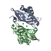 6ucyC  3vsyS S: Starting model for refinement C: citing same article ( |
|---|---|
| Similar structure data |


-Links
 PDBj
PDBj

- Assembly
| Deposited unit | 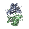
| ||||||||
|---|---|---|---|---|---|---|---|---|---|
| 1 |
| ||||||||
| Unit cell |
|
-Components
| #1: Protein | Mass: 14548.501 Da / Num. of mol.: 2 Source method: isolated from a genetically manipulated source Source: (gene. exp.) Pseudomonas putida (bacteria) / Gene: ksi / Production host: #2: Chemical | 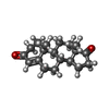  Mass: 286.409 Da / Num. of mol.: 2 / Source method: obtained synthetically / Formula: C19H26O2 / Feature type: SUBJECT OF INVESTIGATION Mass: 286.409 Da / Num. of mol.: 2 / Source method: obtained synthetically / Formula: C19H26O2 / Feature type: SUBJECT OF INVESTIGATION#3: Chemical |   Mass: 35.453 Da / Num. of mol.: 2 / Source method: obtained synthetically / Formula: Cl Mass: 35.453 Da / Num. of mol.: 2 / Source method: obtained synthetically / Formula: Cl#4: Chemical | ChemComp-MG / |   Mass: 24.305 Da / Num. of mol.: 1 / Source method: obtained synthetically / Formula: Mg Mass: 24.305 Da / Num. of mol.: 1 / Source method: obtained synthetically / Formula: Mg#5: Water | ChemComp-HOH / |  Mass: 18.015 Da / Num. of mol.: 120 / Source method: isolated from a natural source / Formula: H2O Mass: 18.015 Da / Num. of mol.: 120 / Source method: isolated from a natural source / Formula: H2OHas ligand of interest | Y | |
|---|
-Experimental details
-Experiment
| Experiment | Method: X-RAY DIFFRACTION / Number of used crystals: 1 |
|---|
- Sample preparation
| Crystal | Density Matthews: 2.31 Å3/Da / Density % sol: 46.69 % |
|---|---|
| Crystal grow | Temperature: 293 K / Method: vapor diffusion, hanging drop / pH: 7.2 / Details: 17-23 % PEG 3350, 0.2 M MAGNESIUM CHLORIDE / Temp details: room temperature |
-Data collection
| Diffraction | Mean temperature: 280 K / Serial crystal experiment: N | ||||||||||||||||||||||||||||||
|---|---|---|---|---|---|---|---|---|---|---|---|---|---|---|---|---|---|---|---|---|---|---|---|---|---|---|---|---|---|---|---|
| Diffraction source | Source: SYNCHROTRON / Site: SSRL / Beamline: BL9-2 / Wavelength: 0.886 Å | ||||||||||||||||||||||||||||||
| Detector | Type: DECTRIS PILATUS 6M / Detector: PIXEL / Date: May 23, 2016 | ||||||||||||||||||||||||||||||
| Radiation | Protocol: SINGLE WAVELENGTH / Monochromatic (M) / Laue (L): M / Scattering type: x-ray | ||||||||||||||||||||||||||||||
| Radiation wavelength | Wavelength: 0.886 Å / Relative weight: 1 | ||||||||||||||||||||||||||||||
| Reflection | Resolution: 1.45→37.19 Å / Num. obs: 46456 / % possible obs: 99.8 % / Redundancy: 5.5 % / Biso Wilson estimate: 19.01 Å2 / CC1/2: 0.999 / Rmerge(I) obs: 0.048 / Rpim(I) all: 0.023 / Rrim(I) all: 0.054 / Net I/σ(I): 18 / Num. measured all: 256187 / Scaling rejects: 35 | ||||||||||||||||||||||||||||||
| Reflection shell | Diffraction-ID: 1
|
- Processing
| Software |
| ||||||||||||||||||||||||||||||||||||||||||||||||||||||||||||||||||||||||||||||||||||||||||||||||||||||
|---|---|---|---|---|---|---|---|---|---|---|---|---|---|---|---|---|---|---|---|---|---|---|---|---|---|---|---|---|---|---|---|---|---|---|---|---|---|---|---|---|---|---|---|---|---|---|---|---|---|---|---|---|---|---|---|---|---|---|---|---|---|---|---|---|---|---|---|---|---|---|---|---|---|---|---|---|---|---|---|---|---|---|---|---|---|---|---|---|---|---|---|---|---|---|---|---|---|---|---|---|---|---|---|
| Refinement | Method to determine structure: MOLECULAR REPLACEMENT Starting model: 3VSY Resolution: 1.4507→34.6595 Å / SU ML: 0.16 / Cross valid method: THROUGHOUT / σ(F): 1.33 / Phase error: 17.33
| ||||||||||||||||||||||||||||||||||||||||||||||||||||||||||||||||||||||||||||||||||||||||||||||||||||||
| Solvent computation | Shrinkage radii: 0.9 Å / VDW probe radii: 1.11 Å | ||||||||||||||||||||||||||||||||||||||||||||||||||||||||||||||||||||||||||||||||||||||||||||||||||||||
| Displacement parameters | Biso max: 109.99 Å2 / Biso mean: 33.3043 Å2 / Biso min: 10.75 Å2 | ||||||||||||||||||||||||||||||||||||||||||||||||||||||||||||||||||||||||||||||||||||||||||||||||||||||
| Refinement step | Cycle: final / Resolution: 1.4507→34.6595 Å
| ||||||||||||||||||||||||||||||||||||||||||||||||||||||||||||||||||||||||||||||||||||||||||||||||||||||
| Refine LS restraints |
| ||||||||||||||||||||||||||||||||||||||||||||||||||||||||||||||||||||||||||||||||||||||||||||||||||||||
| LS refinement shell | Refine-ID: X-RAY DIFFRACTION / Rfactor Rfree error: 0
|
