 Movie
Movie Controller

[English] 日本語
 Yorodumi
- PDB-6ty2: CT PART CRYSTAL STRUCTURE OF THE RYMV-ENCODED VIRAL RNA SILENCING... -
+ Open data

- Basic information
| Entry | Database: PDB / ID: 6ty2 | ||||||
|---|---|---|---|---|---|---|---|
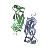
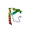
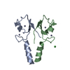
| Title | CT PART CRYSTAL STRUCTURE OF THE RYMV-ENCODED VIRAL RNA SILENCING SUPPRESSOR P1 | ||||||
 Components Components | p1 | ||||||
 Keywords Keywords | RNA BINDING PROTEIN / ZINC FINGERS / RNA SILENCING SUPPRESSOR | ||||||
| Function / homology | metal ion binding / p1 Function and homology information Function and homology information | ||||||
| Biological species |  Rice yellow mottle virus Rice yellow mottle virus | ||||||
| Method |  X-RAY DIFFRACTION / SYNCHROTRON / SAD / Resolution: 1.98 Å X-RAY DIFFRACTION / SYNCHROTRON / SAD / Resolution: 1.98 Å | ||||||
 Authors Authors | Vignols, F. / Hoh, F. | ||||||
 Citation Citation | Journal: J.Mol.Biol. / Year: 2022 Title: A Flexible and Original Architecture of Two Unrelated Zinc Fingers Underlies the Role of the Multitask P1 in RYMV Spread. Authors: Poignavent, V. / Hoh, F. / Terral, G. / Yang, Y. / Gillet, F.X. / Kim, J.H. / Allemand, F. / Lacombe, E. / Brugidou, C. / Cianferani, S. / Demene, H. / Vignols, F. #1: Journal: J. Mol. Biol. / Year: 2013Title: The RYMV-encoded viral suppressor of RNA silencing P1 is a zinc-binding protein with redox-dependent flexibility. Authors: Gillet, F.X. / Cattoni, D.I. / Petiot-Becard, S. / Delalande, F. / Poignavent, V. / Brizard, J.P. / Bessin, Y. / Dorsselaer, A.V. / Declerck, N. / Sanglier-Cianferani, S. / Brugidou, C. / Vignols, F. #2: Journal: Biomol NMR Assign / Year: 2019Title: NMR chemical shift backbone assignment of the viral protein P1 encoded by the African Rice Yellow Mottle Virus. Authors: Yang, Y. / Poignavent, V. / Gillet, F.X. / Vignols, F. / Demene, H. | ||||||
| History |
|
- Structure visualization
| Structure viewer | Molecule: MolmilJmol/JSmol |
|---|
- Downloads & links
-Download
| PDBx/mmCIF format | 6ty2.cif.gz | 35 KB | Display | PDBx/mmCIF format |
|---|---|---|---|---|
| PDB format | pdb6ty2.ent.gz | 22.8 KB | Display | PDB format |
| PDBx/mmJSON format | 6ty2.json.gz | Tree view | PDBx/mmJSON format | |
| Others |  Other downloads Other downloads |
-Validation report
| Arichive directory | https://data.pdbj.org/pub/pdb/validation_reports/ty/6ty2ftp://data.pdbj.org/pub/pdb/validation_reports/ty/6ty2 | HTTPS FTP |
|---|
-Related structure data


-Links
 PDBj
PDBj
- Assembly
| Deposited unit | 
| ||||||||
|---|---|---|---|---|---|---|---|---|---|
| 1 |
| ||||||||
| Unit cell |
|
-Components
| #1: Protein | Mass: 6435.082 Da / Num. of mol.: 2 Source method: isolated from a genetically manipulated source Source: (gene. exp.) Rice yellow mottle virus / Production host:  #2: Chemical |   Mass: 65.409 Da / Num. of mol.: 2 / Source method: obtained synthetically / Formula: Zn Mass: 65.409 Da / Num. of mol.: 2 / Source method: obtained synthetically / Formula: Zn#3: Water | ChemComp-HOH / |  Mass: 18.015 Da / Num. of mol.: 48 / Source method: isolated from a natural source / Formula: H2O Mass: 18.015 Da / Num. of mol.: 48 / Source method: isolated from a natural source / Formula: H2OHas ligand of interest | N | |
|---|
-Experimental details
-Experiment
| Experiment | Method: X-RAY DIFFRACTION / Number of used crystals: 1 |
|---|
- Sample preparation
| Crystal | Density Matthews: 2.01 Å3/Da / Density % sol: 38.87 % |
|---|---|
| Crystal grow | Temperature: 293 K / Method: vapor diffusion, sitting drop Details: 0.1 M tri-Sodium citrate pH 5.6, 20 %(v/v) Isopropanol and 20 %(w/v) PEG 4000. |
-Data collection
| Diffraction | Mean temperature: 100 K / Serial crystal experiment: N |
|---|---|
| Diffraction source | Source: SYNCHROTRON / Site: ESRF  / Beamline: ID23-1 / Wavelength: 1.253 Å / Beamline: ID23-1 / Wavelength: 1.253 Å |
| Detector | Type: DECTRIS PILATUS 6M / Detector: PIXEL / Date: Jul 1, 2013 |
| Radiation | Protocol: SINGLE WAVELENGTH / Monochromatic (M) / Laue (L): M / Scattering type: x-ray |
| Radiation wavelength | Wavelength: 1.253 Å / Relative weight: 1 |
| Reflection | Resolution: 1.68→41.29 Å / Num. obs: 11192 / % possible obs: 99 % / Redundancy: 7 % / CC1/2: 1 / CC star: 0.08 / Rmerge(I) obs: 0.01 / Rrim(I) all: 0.015 / Net I/σ(I): 7 |
| Reflection shell | Resolution: 1.68→1.77 Å / Redundancy: 3 % / Rmerge(I) obs: 0.9 / Mean I/σ(I) obs: 1.3 / Num. unique obs: 1535 / Rpim(I) all: 0.6 / % possible all: 95 |
- Processing
| Software |
| ||||||||||||||||||||||||
|---|---|---|---|---|---|---|---|---|---|---|---|---|---|---|---|---|---|---|---|---|---|---|---|---|---|
| Refinement | Method to determine structure: SAD / Resolution: 1.98→37.368 Å / SU ML: 0.21 / Cross valid method: FREE R-VALUE / σ(F): 1.35 / Phase error: 25.26
| ||||||||||||||||||||||||
| Solvent computation | Shrinkage radii: 0.9 Å / VDW probe radii: 1.11 Å | ||||||||||||||||||||||||
| Refinement step | Cycle: LAST / Resolution: 1.98→37.368 Å
| ||||||||||||||||||||||||
| Refine LS restraints |
| ||||||||||||||||||||||||
| LS refinement shell | Resolution: 1.9805→2.4951 Å
|
