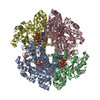
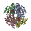
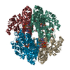
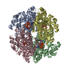
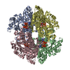
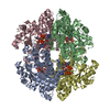
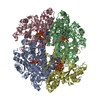
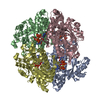
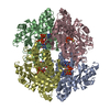
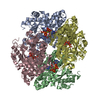
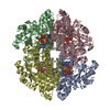
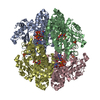
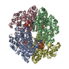
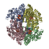
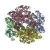
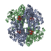
[English] 日本語
Yorodumi
- PDB-6txf: Crystal structure of tetrameric human D137N-SAMHD1 (residues 109-... -

+
Open data


ID or keywords:

Loading...

-
Basic information

Entry
Database: PDB / ID: 6txf
TitleCrystal structure of tetrameric human D137N-SAMHD1 (residues 109-626) with XTP, dAMPNPP and Mn
ComponentsDeoxynucleoside triphosphate triphosphohydrolase SAMHD1
KeywordsHYDROLASE / triphosphohydrolase / metallo-enzyme / binuclear / HD
Function / homology
Function and homology information


Nucleotide catabolism / Hydrolases; Acting on ester bonds; Triphosphoric-monoester hydrolases / deoxynucleoside triphosphate hydrolase activity / triphosphoric monoester hydrolase activity / dGTP binding / dATP catabolic process / dGTPase activity / tetraspanin-enriched microdomain / DNA strand resection involved in replication fork processing / deoxyribonucleotide catabolic process ...Nucleotide catabolism / Hydrolases; Acting on ester bonds; Triphosphoric-monoester hydrolases / deoxynucleoside triphosphate hydrolase activity / triphosphoric monoester hydrolase activity / dGTP binding / dATP catabolic process / dGTPase activity / tetraspanin-enriched microdomain / DNA strand resection involved in replication fork processing / deoxyribonucleotide catabolic process / dGTP catabolic process / negative regulation of type I interferon-mediated signaling pathway / regulation of innate immune response / somatic hypermutation of immunoglobulin genes / RNA nuclease activity / double-strand break repair via homologous recombination / Interferon alpha/beta signaling / site of double-strand break / single-stranded DNA binding / protein homotetramerization / defense response to virus / nucleic acid binding / immune response / innate immune response / DNA damage response / GTP binding / RNA binding / zinc ion binding / nucleoplasm / identical protein binding / nucleus / plasma membrane
Similarity search - Function
HD domain profile. / HD domain / HD domain / SAM domain (Sterile alpha motif) / SAM domain profile. / Metal dependent phosphohydrolases with conserved 'HD' motif. / Sterile alpha motif. / HD/PDEase domain / Sterile alpha motif domain / Sterile alpha motif/pointed domain superfamily
Similarity search - Domain/homology
Chem-CZF / Chem-DZ4 / : / : / Deoxynucleoside triphosphate triphosphohydrolase SAMHD1
Similarity search - Component
Biological speciesHomo sapiens (human)
MethodX-RAY DIFFRACTION / SYNCHROTRON / MOLECULAR REPLACEMENT / molecular replacement / Resolution: 2.25 Å
AuthorsMorris, E.R. / Kunzelmann, S. / Caswell, S.J. / Arnold, L.H. / Purkiss, A.G. / Kelly, G. / Taylor, I.A.
Funding support United Kingdom, 4items
OrganizationGrant numberCountry
Wellcome Trust108014/Z/15/Z United Kingdom
Wellcome TrustFC001029 United Kingdom
Medical Research Council (MRC, United Kingdom)FC001029 United Kingdom
Cancer Research UKFC001029 United Kingdom
CitationJournal: Nat Commun / Year: 2020
Title: Crystal structures of SAMHD1 inhibitor complexes reveal the mechanism of water-mediated dNTP hydrolysis.
Authors: Morris, E.R. / Caswell, S.J. / Kunzelmann, S. / Arnold, L.H. / Purkiss, A.G. / Kelly, G. / Taylor, I.A.
History
DepositionJan 14, 2020Deposition site: PDBE / Processing site: PDBE
Revision 1.0Jun 24, 2020Provider: repository / Type: Initial release
Revision 1.1Jul 8, 2020Group: Database references / Category: citation / citation_author
Item: _citation.journal_volume / _citation.page_first ..._citation.journal_volume / _citation.page_first / _citation.page_last / _citation.pdbx_database_id_PubMed / _citation.title / _citation_author.identifier_ORCID / _citation_author.name
Revision 1.2Jan 24, 2024Group: Data collection / Database references ...Data collection / Database references / Derived calculations / Refinement description / Structure summary
Category: chem_comp / chem_comp_atom ...chem_comp / chem_comp_atom / chem_comp_bond / database_2 / pdbx_initial_refinement_model / pdbx_struct_conn_angle / struct_conn / struct_ncs_dom_lim
Item: _chem_comp.pdbx_synonyms / _database_2.pdbx_DOI ..._chem_comp.pdbx_synonyms / _database_2.pdbx_DOI / _database_2.pdbx_database_accession / _pdbx_struct_conn_angle.ptnr1_auth_asym_id / _pdbx_struct_conn_angle.ptnr1_auth_comp_id / _pdbx_struct_conn_angle.ptnr1_auth_seq_id / _pdbx_struct_conn_angle.ptnr1_label_asym_id / _pdbx_struct_conn_angle.ptnr1_label_atom_id / _pdbx_struct_conn_angle.ptnr1_label_comp_id / _pdbx_struct_conn_angle.ptnr1_label_seq_id / _pdbx_struct_conn_angle.ptnr2_auth_asym_id / _pdbx_struct_conn_angle.ptnr2_auth_comp_id / _pdbx_struct_conn_angle.ptnr2_auth_seq_id / _pdbx_struct_conn_angle.ptnr2_label_asym_id / _pdbx_struct_conn_angle.ptnr2_label_atom_id / _pdbx_struct_conn_angle.ptnr2_label_comp_id / _pdbx_struct_conn_angle.ptnr3_auth_asym_id / _pdbx_struct_conn_angle.ptnr3_auth_comp_id / _pdbx_struct_conn_angle.ptnr3_auth_seq_id / _pdbx_struct_conn_angle.ptnr3_label_asym_id / _pdbx_struct_conn_angle.ptnr3_label_atom_id / _pdbx_struct_conn_angle.ptnr3_label_comp_id / _pdbx_struct_conn_angle.ptnr3_label_seq_id / _pdbx_struct_conn_angle.value / _struct_conn.pdbx_dist_value / _struct_conn.ptnr1_auth_asym_id / _struct_conn.ptnr1_auth_comp_id / _struct_conn.ptnr1_auth_seq_id / _struct_conn.ptnr1_label_asym_id / _struct_conn.ptnr1_label_atom_id / _struct_conn.ptnr1_label_comp_id / _struct_conn.ptnr1_label_seq_id / _struct_conn.ptnr2_auth_asym_id / _struct_conn.ptnr2_auth_comp_id / _struct_conn.ptnr2_auth_seq_id / _struct_conn.ptnr2_label_asym_id / _struct_conn.ptnr2_label_atom_id / _struct_conn.ptnr2_label_comp_id / _struct_ncs_dom_lim.beg_auth_comp_id / _struct_ncs_dom_lim.beg_label_asym_id / _struct_ncs_dom_lim.beg_label_comp_id / _struct_ncs_dom_lim.beg_label_seq_id / _struct_ncs_dom_lim.end_auth_comp_id / _struct_ncs_dom_lim.end_label_asym_id / _struct_ncs_dom_lim.end_label_comp_id / _struct_ncs_dom_lim.end_label_seq_id

-
Structure visualization

Structure viewerMolecule:
MolmilJmol/JSmol

Downloads & links

-
Assembly

Deposited unit
A: Deoxynucleoside triphosphate triphosphohydrolase SAMHD1
B: Deoxynucleoside triphosphate triphosphohydrolase SAMHD1
hetero molecules


Theoretical massNumber of molelcules
Total (without water)123,86616
Polymers120,4162
Non-polymers3,45014
Water1,62190
1
A: Deoxynucleoside triphosphate triphosphohydrolase SAMHD1
B: Deoxynucleoside triphosphate triphosphohydrolase SAMHD1
hetero molecules

A: Deoxynucleoside triphosphate triphosphohydrolase SAMHD1
B: Deoxynucleoside triphosphate triphosphohydrolase SAMHD1
hetero molecules


Theoretical massNumber of molelcules
Total (without water)247,73232
Polymers240,8314
Non-polymers6,90128
Water724
TypeNameSymmetry operationNumber
identity operation1_555x,y,z1
crystal symmetry operation7_555y,x,-z1
Buried area23220 Å2
ΔGint-138 kcal/mol
Surface area67290 Å2
MethodPISA
Unit cell
Length a, b, c (Å)104.978, 104.978, 195.516
Angle α, β, γ (deg.)90.000, 90.000, 90.000
Int Tables number96
Space group name H-MP43212
Noncrystallographic symmetry (NCS)NCS domain:
IDEns-IDDetails
11A
21B

NCS domain segments:

Component-ID: _ / Ens-ID: 1 / Beg auth comp-ID: THR / Beg label comp-ID: THR / End auth comp-ID: ASN / End label comp-ID: ASN / Refine code: _ / Auth seq-ID: 114 - 599 / Label seq-ID: 8 - 493

Dom-IDAuth asym-IDLabel asym-ID
1AA
2BB

-
Components

-
Protein , 1 types, 2 molecules AB

#1: Protein Deoxynucleoside triphosphate triphosphohydrolase SAMHD1 / dNTPase / Dendritic cell-derived IFNG-induced protein / DCIP / Monocyte protein 5 / MOP-5 / SAM ...dNTPase / Dendritic cell-derived IFNG-induced protein / DCIP / Monocyte protein 5 / MOP-5 / SAM domain and HD domain-containing protein 1 / hSAMHD1


Mass: 60207.816 Da / Num. of mol.: 2 / Mutation: D137N
Source method: isolated from a genetically manipulated source
Source: (gene. exp.) Homo sapiens (human) / Gene: SAMHD1, MOP5 / Production host: Escherichia coli BL21(DE3) (bacteria) / Variant (production host): Rosetta2
References: UniProt: Q9Y3Z3, Hydrolases; Acting on ester bonds; Triphosphoric-monoester hydrolases

-
Non-polymers , 5 types, 104 molecules

#2: Chemical ChemComp-FE / FE (III) ION


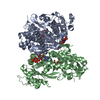
Mass: 55.845 Da / Num. of mol.: 2 / Source method: obtained synthetically / Formula: Fe / Feature type: SUBJECT OF INVESTIGATION
#3: Chemical
ChemComp-MN / MANGANESE (II) ION


Mass: 54.938 Da / Num. of mol.: 6 / Source method: obtained synthetically / Formula: Mn / Feature type: SUBJECT OF INVESTIGATION
#4: Chemical
ChemComp-DZ4 / 2'-deoxy-5'-O-[(R)-hydroxy{[(R)-hydroxy(phosphonooxy)phosphoryl]amino}phosphoryl]adenosine


Mass: 490.197 Da / Num. of mol.: 4 / Source method: obtained synthetically / Formula: C10H17N6O11P3 / Feature type: SUBJECT OF INVESTIGATION
#5: Chemical ChemComp-CZF / [[(2~{R},3~{S},4~{R},5~{R})-5-[2,6-bis(oxidanylidene)-3~{H}-purin-9-yl]-3,4-bis(oxidanyl)oxolan-2-yl]methoxy-oxidanyl-phosphoryl] phosphono hydrogen phosphate / xanthosine triphosphate


Mass: 524.165 Da / Num. of mol.: 2 / Source method: obtained synthetically / Formula: C10H15N4O15P3 / Feature type: SUBJECT OF INVESTIGATION
#6: Water ChemComp-HOH / water


Mass: 18.015 Da / Num. of mol.: 90 / Source method: isolated from a natural source / Formula: H2O

-
Details

Has ligand of interestY

-
Experimental details

-
Experiment

ExperimentMethod: X-RAY DIFFRACTION / Number of used crystals: 1

-
Sample preparation

CrystalDensity Matthews: 2.24 Å3/Da / Density % sol: 45.12 %
Crystal growTemperature: 293 K / Method: vapor diffusion, sitting drop / pH: 6 / Details: 0.1 M Bistris methane-HCl pH 6, 14% (w/v) PEG 3350

-
Data collection

DiffractionMean temperature: 100 K / Serial crystal experiment: N
Diffraction sourceSource: SYNCHROTRON / Site: Diamond / Beamline: I24 / Wavelength: 0.97866 Å
DetectorType: DECTRIS PILATUS3 6M / Detector: PIXEL / Date: Apr 22, 2017
RadiationProtocol: SINGLE WAVELENGTH / Monochromatic (M) / Laue (L): M / Scattering type: x-ray
Radiation wavelengthWavelength: 0.97866 Å / Relative weight: 1
ReflectionResolution: 2.25→71.59 Å / Num. obs: 51603 / % possible obs: 97.75 % / Redundancy: 16.83 % / Biso Wilson estimate: 37.6 Å2 / CC1/2: 0.998 / Rmerge(I) obs: 0.122 / Rpim(I) all: 0.029 / Rrim(I) all: 0.126 / Net I/σ(I): 14.5
Reflection shell

Diffraction-ID: 1

Resolution (Å)Redundancy (%)Rmerge(I) obsMean I/σ(I) obsNum. unique obsCC1/2Rpim(I) allRrim(I) all% possible all
2.25-2.296.841.2161.82850.6730.4631.30780.13
6.1-71.5818.560.08331.529300.9970.020.085100

-
Phasing

PhasingMethod: molecular replacement
Phasing MRModel details: Phaser MODE: MR_AUTO
Highest resolutionLowest resolution
Rotation7.08 Å71.54 Å
Translation7.08 Å71.54 Å

-
Processing

Software
NameVersionClassificationNB
XDSdata reduction
Aimless0.5.32data scaling
PHASER2.7.17phasing
REFMAC5.8.0158refinement
PDB_EXTRACT3.25data extraction
RefinementMethod to determine structure: MOLECULAR REPLACEMENT
Starting model: 6TX0
Resolution: 2.25→71.51 Å / Cor.coef. Fo:Fc: 0.956 / Cor.coef. Fo:Fc free: 0.933 / SU B: 16.929 / SU ML: 0.188 / SU R Cruickshank DPI: 0.3137 / Cross valid method: THROUGHOUT / σ(F): 0 / ESU R: 0.314 / ESU R Free: 0.224
Details: HYDROGENS HAVE BEEN ADDED IN THE RIDING POSITIONS U VALUES : WITH TLS ADDED
RfactorNum. reflection% reflectionSelection details
Rfree0.2388 2568 5 %RANDOM
Rwork0.1952 ---
obs0.1974 48696 97.42 %-
Solvent computationIon probe radii: 0.8 Å / Shrinkage radii: 0.8 Å / VDW probe radii: 1.2 Å
Displacement parametersBiso max: 157.65 Å2 / Biso mean: 47.142 Å2 / Biso min: 21.5 Å2
Baniso -1Baniso -2Baniso -3
1-0.17 Å2-0 Å2-0 Å2
2--0.17 Å2-0 Å2
3----0.34 Å2
Refinement stepCycle: final / Resolution: 2.25→71.51 Å
ProteinNucleic acidLigandSolventTotal
Num. atoms7748 0 192 90 8030
Biso mean--32.33 32.52 -
Num. residues----958
Refine LS restraints
Refine-IDTypeDev idealDev ideal targetNumber
X-RAY DIFFRACTIONr_bond_refined_d0.0210.0198130
X-RAY DIFFRACTIONr_bond_other_d0.0020.027362
X-RAY DIFFRACTIONr_angle_refined_deg2.0951.95211043
X-RAY DIFFRACTIONr_angle_other_deg1.142.98817083
X-RAY DIFFRACTIONr_dihedral_angle_1_deg5.9425954
X-RAY DIFFRACTIONr_dihedral_angle_2_deg38.70123.955397
X-RAY DIFFRACTIONr_dihedral_angle_3_deg15.242151381
X-RAY DIFFRACTIONr_dihedral_angle_4_deg16.9291553
X-RAY DIFFRACTIONr_chiral_restr0.1220.21190
X-RAY DIFFRACTIONr_gen_planes_refined0.0110.0218913
X-RAY DIFFRACTIONr_gen_planes_other0.0030.021666
Refine LS restraints NCS

Ens-ID: 1 / Number: 31838 / Refine-ID: X-RAY DIFFRACTION / Type: interatomic distance / Rms dev position: 0.06 Å / Weight position: 0.05

Dom-IDAuth asym-ID
1A
2B
LS refinement shellResolution: 2.25→2.308 Å / Rfactor Rfree error: 0 / Total num. of bins used: 20
RfactorNum. reflection% reflection
Rfree0.357 164 -
Rwork0.349 3057 -
all-3221 -
obs--85.17 %
Refinement TLS params.

Method: refined / Refine-ID: X-RAY DIFFRACTION

IDL112)L122)L132)L222)L232)L332)S11 (Å °)S12 (Å °)S13 (Å °)S21 (Å °)S22 (Å °)S23 (Å °)S31 (Å °)S32 (Å °)S33 (Å °)T112)T122)T132)T222)T232)T332)Origin x (Å)Origin y (Å)Origin z (Å)
10.1689-0.07940.0890.23370.03680.22940.0815-0.0579-0.0101-0.0995-0.0157-0.0790.0780.0509-0.06580.15930.02590.00070.0940.01190.150620.6861-13.8989-4.3324
20.29960.01930.16120.1847-0.10590.24340.0821-0.02650.0097-0.23440.0230.08940.1685-0.023-0.10510.3441-0.039-0.09510.01480.00780.0797-6.8982-4.6964-24.836
Refinement TLS group
IDRefine-IDRefine TLS-IDAuth asym-IDAuth seq-ID
1X-RAY DIFFRACTION1A114 - 901
2X-RAY DIFFRACTION2B114 - 901

+
About Yorodumi

-
News

-
Feb 9, 2022. New format data for meta-information of EMDB entries

New format data for meta-information of EMDB entries

  • Version 3 of the EMDB header file is now the official format.
  • The previous official version 1.9 will be removed from the archive.

Related info.:EMDB header

External links:wwPDB to switch to version 3 of the EMDB data model

-
Aug 12, 2020. Covid-19 info

Covid-19 info

URL: https://pdbjlvh1.pdbj.org/emnavi/covid19.php

New page: Covid-19 featured information page in EM Navigator.

Related info.:Covid-19 info / Mar 5, 2020. Novel coronavirus structure data

+
Mar 5, 2020. Novel coronavirus structure data

Novel coronavirus structure data

Related info.:Yorodumi Speices / Aug 12, 2020. Covid-19 info

External links:COVID-19 featured content - PDBj / Molecule of the Month (242):Coronavirus Proteases

+
Jan 31, 2019. EMDB accession codes are about to change! (news from PDBe EMDB page)

EMDB accession codes are about to change! (news from PDBe EMDB page)

  • The allocation of 4 digits for EMDB accession codes will soon come to an end. Whilst these codes will remain in use, new EMDB accession codes will include an additional digit and will expand incrementally as the available range of codes is exhausted. The current 4-digit format prefixed with “EMD-” (i.e. EMD-XXXX) will advance to a 5-digit format (i.e. EMD-XXXXX), and so on. It is currently estimated that the 4-digit codes will be depleted around Spring 2019, at which point the 5-digit format will come into force.
  • The EM Navigator/Yorodumi systems omit the EMD- prefix.

Related info.:Q: What is EMD? / ID/Accession-code notation in Yorodumi/EM Navigator

External links:EMDB Accession Codes are Changing Soon! / Contact to PDBj

+
Jul 12, 2017. Major update of PDB

Major update of PDB

  • wwPDB released updated PDB data conforming to the new PDBx/mmCIF dictionary.
  • This is a major update changing the version number from 4 to 5, and with Remediation, in which all the entries are updated.
  • In this update, many items about electron microscopy experimental information are reorganized (e.g. em_software).
  • Now, EM Navigator and Yorodumi are based on the updated data.

External links:wwPDB Remediation / Enriched Model Files Conforming to OneDep Data Standards Now Available in the PDB FTP Archive

-
Yorodumi

Thousand views of thousand structures

  • Yorodumi is a browser for structure data from EMDB, PDB, SASBDB, etc.
  • This page is also the successor to EM Navigator detail page, and also detail information page/front-end page for Omokage search.
  • The word "yorodu" (or yorozu) is an old Japanese word meaning "ten thousand". "mi" (miru) is to see.

Related info.:EMDB / PDB / SASBDB / Comparison of 3 databanks / Yorodumi Search / Aug 31, 2016. New EM Navigator & Yorodumi / Yorodumi Papers / Jmol/JSmol / Function and homology information / Changes in new EM Navigator and Yorodumi

Read more