 ムービー
ムービー コントローラー


+ データを開く

- 基本情報
基本情報
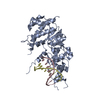
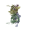
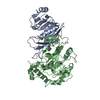
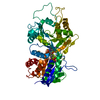
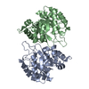
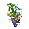
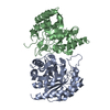
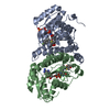
| 登録情報 | データベース: PDB / ID: 6tuw | ||||||
|---|---|---|---|---|---|---|---|
| タイトル | human XPG-DNA, Complex 1 | ||||||
 要素 要素 |
| ||||||
 キーワード キーワード | DNA BINDING PROTEIN / XPG nuclease domain bound to DNA | ||||||
| 機能・相同性 |  機能・相同性情報 機能・相同性情報nucleotide-excision repair complex / base-excision repair, AP site formation / bubble DNA binding / response to UV-C / RNA polymerase II complex binding / response to UV / transcription-coupled nucleotide-excision repair / DNA endonuclease activity / nucleotide-excision repair / enzyme activator activity ...nucleotide-excision repair complex / base-excision repair, AP site formation / bubble DNA binding / response to UV-C / RNA polymerase II complex binding / response to UV / transcription-coupled nucleotide-excision repair / DNA endonuclease activity / nucleotide-excision repair / enzyme activator activity / double-strand break repair via homologous recombination / Dual Incision in GG-NER / Formation of Incision Complex in GG-NER / Dual incision in TC-NER / single-stranded DNA binding / chromosome / double-stranded DNA binding / endonuclease activity / damaged DNA binding / 加水分解酵素; エステル加水分解酵素 / negative regulation of apoptotic process / protein-containing complex binding / protein homodimerization activity / protein-containing complex / nucleoplasm / metal ion binding / nucleus 類似検索 - 分子機能 | ||||||
| 生物種 |  Homo sapiens (ヒト) Homo sapiens (ヒト)unidentified (未定義) | ||||||
| 手法 |  X線回折 / シンクロトロン / 分子置換 / 解像度: 3.5 Å X線回折 / シンクロトロン / 分子置換 / 解像度: 3.5 Å | ||||||
 データ登録者 データ登録者 | Ruiz, F.M. / Fernandez-Tornero, C. | ||||||
 引用 引用 | ジャーナル: Nucleic Acids Res. / 年: 2020 タイトル: The crystal structure of human XPG, the xeroderma pigmentosum group G endonuclease, provides insight into nucleotide excision DNA repair. 著者: Gonzalez-Corrochano, R. / Ruiz, F.M. / Taylor, N.M.I. / Huecas, S. / Drakulic, S. / Spinola-Amilibia, M. / Fernandez-Tornero, C. | ||||||
| 履歴 |
|
- 構造の表示
構造の表示
| 構造ビューア | 分子: MolmilJmol/JSmol |
|---|
- ダウンロードとリンク
-ダウンロード
| PDBx/mmCIF形式 | 6tuw.cif.gz | 107.5 KB | 表示 | PDBx/mmCIF形式 |
|---|---|---|---|---|
| PDB形式 | pdb6tuw.ent.gz | 65.1 KB | 表示 | PDB形式 |
| PDBx/mmJSON形式 | 6tuw.json.gz | ツリー表示 | PDBx/mmJSON形式 | |
| その他 |  その他のダウンロード その他のダウンロード |
-検証レポート
| 文書・要旨 | 6tuw_validation.pdf.gz | 447.2 KB | 表示 | wwPDB検証レポート |
|---|---|---|---|---|
| 文書・詳細版 | 6tuw_full_validation.pdf.gz | 451.5 KB | 表示 | |
| XML形式データ | 6tuw_validation.xml.gz | 13.8 KB | 表示 | |
| CIF形式データ | 6tuw_validation.cif.gz | 17.6 KB | 表示 | |
| アーカイブディレクトリ | https://data.pdbj.org/pub/pdb/validation_reports/tu/6tuwftp://data.pdbj.org/pub/pdb/validation_reports/tu/6tuw | HTTPS FTP |
-関連構造データ


-リンク
 PDBj
PDBj


- 集合体
集合体
| 登録構造単位 | 
| ||||||||||||
|---|---|---|---|---|---|---|---|---|---|---|---|---|---|
| 1 |
| ||||||||||||
| 単位格子 |
|
-要素
| #1: タンパク質 | 分子量: 40789.887 Da / 分子数: 1 / 由来タイプ: 組換発現 / 由来: (組換発現) Homo sapiens (ヒト) / 遺伝子: ERCC5, ERCM2, XPG, XPGC / 発現宿主:  参照: UniProt: P28715, 加水分解酵素; エステル加水分解酵素 |
|---|---|
| #2: DNA鎖 | 分子量: 3364.208 Da / 分子数: 1 / 由来タイプ: 合成 / 由来: (合成) unidentified (未定義) |
| #3: DNA鎖 | 分子量: 3358.211 Da / 分子数: 1 / 由来タイプ: 合成 / 由来: (合成) unidentified (未定義) |
-実験情報
-実験
| 実験 | 手法: X線回折 / 使用した結晶の数: 1 |
|---|
- 試料調製
試料調製
| 結晶 | マシュー密度: 2.62 Å3/Da / 溶媒含有率: 53.05 % |
|---|---|
| 結晶化 | 温度: 295 K / 手法: 蒸気拡散法, シッティングドロップ法 / 詳細: 5% PEG 3350, 50 mM Na Citrate pH 4.0 |
-データ収集
| 回折 | 平均測定温度: 100 K / Serial crystal experiment: N |
|---|---|
| 放射光源 | 由来: シンクロトロン / サイト: ESRF  / ビームライン: ID29 / 波長: 0.9795 Å / ビームライン: ID29 / 波長: 0.9795 Å |
| 検出器 | タイプ: DECTRIS PILATUS 6M-F / 検出器: PIXEL / 日付: 2015年2月1日 |
| 放射 | プロトコル: SINGLE WAVELENGTH / 単色(M)・ラウエ(L): M / 散乱光タイプ: x-ray |
| 放射波長 | 波長: 0.9795 Å / 相対比: 1 |
| 反射 | 解像度: 3.5→45.79 Å / Num. obs: 6627 / % possible obs: 99.8 % / 冗長度: 15.9 % / Biso Wilson estimate: 145.35 Å2 / CC1/2: 0.998 / Net I/σ(I): 11.1 |
| 反射 シェル | 解像度: 3.5→3.625 Å / Num. unique obs: 1536 / CC1/2: 0.827 |
- 解析
解析
| ソフトウェア |
| ||||||||||||||||||||||||
|---|---|---|---|---|---|---|---|---|---|---|---|---|---|---|---|---|---|---|---|---|---|---|---|---|---|
| 精密化 | 構造決定の手法: 分子置換 開始モデル: 6TUR 解像度: 3.5→45.79 Å / SU ML: 0.3407 / 交差検証法: FREE R-VALUE / σ(F): 1.34 / 位相誤差: 39.6614
| ||||||||||||||||||||||||
| 溶媒の処理 | 減衰半径: 0.9 Å / VDWプローブ半径: 1.11 Å | ||||||||||||||||||||||||
| 原子変位パラメータ | Biso mean: 212.3 Å2 | ||||||||||||||||||||||||
| 精密化ステップ | サイクル: LAST / 解像度: 3.5→45.79 Å
| ||||||||||||||||||||||||
| 拘束条件 |
| ||||||||||||||||||||||||
| LS精密化 シェル |
|
