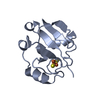
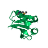
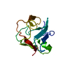
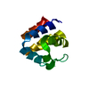
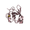
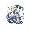
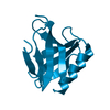
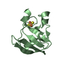
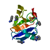
[English] 日本語
Yorodumi
- PDB-6tne: Crystal structure of receiver domain from Hybrid Histidine Kinase CckA -

+
Open data


ID or keywords:

Loading...

-
Basic information

Entry
Database: PDB / ID: 6tne
TitleCrystal structure of receiver domain from Hybrid Histidine Kinase CckA
ComponentsHistidine kinase
KeywordsTRANSFERASE / Cyclic di-GMP / second messenger / hybride histidine kinase / CcKA / phosphorylation
Function / homology
Function and homology information


histidine kinase / phosphorelay sensor kinase activity / nucleotide binding / membrane
Similarity search - Function
His Kinase A (phospho-acceptor) domain / His Kinase A (phosphoacceptor) domain / Signal transduction histidine kinase, dimerisation/phosphoacceptor domain / Signal transduction histidine kinase, dimerisation/phosphoacceptor domain superfamily / Signal transduction histidine kinase-related protein, C-terminal / Histidine kinase domain / Histidine kinase domain profile. / Response regulator receiver domain / cheY-homologous receiver domain / Signal transduction response regulator, receiver domain ...His Kinase A (phospho-acceptor) domain / His Kinase A (phosphoacceptor) domain / Signal transduction histidine kinase, dimerisation/phosphoacceptor domain / Signal transduction histidine kinase, dimerisation/phosphoacceptor domain superfamily / Signal transduction histidine kinase-related protein, C-terminal / Histidine kinase domain / Histidine kinase domain profile. / Response regulator receiver domain / cheY-homologous receiver domain / Signal transduction response regulator, receiver domain / Response regulatory domain profile. / CheY-like superfamily / Histidine kinase-, DNA gyrase B-, and HSP90-like ATPase / Histidine kinase-like ATPases / Histidine kinase/HSP90-like ATPase / Histidine kinase/HSP90-like ATPase superfamily
Similarity search - Domain/homology
Biological speciesCaulobacter vibrioides (bacteria)
MethodX-RAY DIFFRACTION / SYNCHROTRON / MOLECULAR REPLACEMENT / molecular replacement / Resolution: 1.25 Å
AuthorsDubey, B.N. / Bruederlin, M. / Schirmer, T.
Funding support Switzerland, 1items
OrganizationGrant numberCountry
Swiss National Science Foundation31003A-166652 Switzerland
CitationJournal: to be published
Title: Crystal structure of receiver domain from cyclic-di-GMP controlled hybrid histidine kinase
Authors: Dubey, B.N. / Bruederlin, M. / Boehm, R. / Jenal, U. / Hiller, S. / Schirmer, T.
History
DepositionDec 6, 2019Deposition site: PDBE / Processing site: PDBE
Revision 1.0Dec 16, 2020Provider: repository / Type: Initial release
Revision 2.0Oct 5, 2022Group: Advisory / Atomic model ...Advisory / Atomic model / Data collection / Database references / Derived calculations / Non-polymer description / Other / Polymer sequence / Refinement description / Source and taxonomy / Structure summary
Category: atom_site / atom_site_anisotrop ...atom_site / atom_site_anisotrop / atom_sites / chem_comp / database_2 / entity / entity_poly / entity_poly_seq / entity_src_gen / pdbx_contact_author / pdbx_distant_solvent_atoms / pdbx_nonpoly_scheme / pdbx_poly_seq_scheme / pdbx_refine_tls / pdbx_refine_tls_group / pdbx_struct_sheet_hbond / pdbx_unobs_or_zero_occ_residues / pdbx_validate_close_contact / pdbx_validate_rmsd_angle / pdbx_validate_rmsd_bond / pdbx_validate_symm_contact / refine / refine_hist / refine_ls_restr / refine_ls_shell / reflns / reflns_shell / software / struct_conf / struct_mon_prot_cis / struct_ref / struct_ref_seq / struct_ref_seq_dif / struct_sheet_range
Item: _atom_sites.fract_transf_matrix[1][3] / _chem_comp.formula ..._atom_sites.fract_transf_matrix[1][3] / _chem_comp.formula / _chem_comp.formula_weight / _chem_comp.id / _chem_comp.mon_nstd_flag / _chem_comp.name / _chem_comp.type / _database_2.pdbx_DOI / _database_2.pdbx_database_accession / _entity.formula_weight / _entity.pdbx_description / _entity.pdbx_ec / _entity.pdbx_number_of_molecules / _entity_poly.pdbx_seq_one_letter_code / _entity_poly.pdbx_seq_one_letter_code_can / _entity_src_gen.pdbx_end_seq_num / _pdbx_contact_author.id / _pdbx_struct_sheet_hbond.range_1_auth_comp_id / _pdbx_struct_sheet_hbond.range_1_auth_seq_id / _pdbx_struct_sheet_hbond.range_1_label_comp_id / _pdbx_struct_sheet_hbond.range_1_label_seq_id / _pdbx_struct_sheet_hbond.range_2_label_seq_id / _pdbx_validate_symm_contact.auth_atom_id_1 / _pdbx_validate_symm_contact.auth_comp_id_1 / _pdbx_validate_symm_contact.auth_seq_id_1 / _pdbx_validate_symm_contact.auth_seq_id_2 / _pdbx_validate_symm_contact.dist / _pdbx_validate_symm_contact.site_symmetry_2 / _refine.B_iso_max / _refine.B_iso_mean / _refine.B_iso_min / _refine.aniso_B[1][1] / _refine.aniso_B[1][2] / _refine.aniso_B[1][3] / _refine.aniso_B[2][2] / _refine.aniso_B[2][3] / _refine.aniso_B[3][3] / _refine.correlation_coeff_Fo_to_Fc / _refine.correlation_coeff_Fo_to_Fc_free / _refine.details / _refine.ls_R_factor_R_free / _refine.ls_R_factor_R_work / _refine.ls_R_factor_obs / _refine.ls_d_res_low / _refine.ls_number_reflns_R_free / _refine.ls_number_reflns_R_work / _refine.ls_number_reflns_obs / _refine.ls_percent_reflns_R_free / _refine.overall_SU_B / _refine.overall_SU_ML / _refine.overall_SU_R_Cruickshank_DPI / _refine.pdbx_ls_sigma_F / _refine.pdbx_overall_ESU_R / _refine.pdbx_overall_ESU_R_Free / _refine.pdbx_overall_phase_error / _refine.pdbx_solvent_ion_probe_radii / _refine.pdbx_solvent_shrinkage_radii / _refine.pdbx_solvent_vdw_probe_radii / _refine.pdbx_stereochemistry_target_values / _refine.solvent_model_details / _refine_hist.d_res_low / _refine_hist.number_atoms_solvent / _refine_hist.number_atoms_total / _refine_hist.pdbx_B_iso_mean_solvent / _refine_hist.pdbx_number_atoms_protein / _refine_hist.pdbx_number_residues_total / _reflns.B_iso_Wilson_estimate / _reflns.d_resolution_low / _reflns.number_obs / _reflns.pdbx_Rmerge_I_obs / _reflns.pdbx_Rpim_I_all / _reflns.pdbx_Rrim_I_all / _reflns.pdbx_netI_over_sigmaI / _reflns.pdbx_number_measured_all / _reflns.pdbx_redundancy / _reflns.pdbx_scaling_rejects / _reflns.percent_possible_obs / _software.classification / _software.contact_author / _software.contact_author_email / _software.language / _software.location / _software.name / _software.type / _software.version / _struct_conf.beg_label_seq_id / _struct_conf.end_auth_comp_id / _struct_conf.end_auth_seq_id / _struct_conf.end_label_comp_id / _struct_conf.end_label_seq_id / _struct_conf.pdbx_PDB_helix_length / _struct_mon_prot_cis.label_seq_id / _struct_mon_prot_cis.pdbx_label_seq_id_2 / _struct_mon_prot_cis.pdbx_omega_angle / _struct_ref.pdbx_align_begin / _struct_ref.pdbx_seq_one_letter_code / _struct_ref_seq.db_align_beg / _struct_ref_seq.db_align_end / _struct_ref_seq.pdbx_auth_seq_align_beg / _struct_ref_seq.pdbx_auth_seq_align_end / _struct_ref_seq.seq_align_beg / _struct_ref_seq.seq_align_end / _struct_sheet_range.beg_label_seq_id / _struct_sheet_range.end_label_seq_id
Description: Model completeness / Provider: author / Type: Coordinate replacement
Revision 2.1May 1, 2024Group: Data collection / Refinement description
Category: chem_comp_atom / chem_comp_bond / pdbx_initial_refinement_model

-
Structure visualization

Structure viewerMolecule:
MolmilJmol/JSmol

Downloads & links

-
Assembly

Deposited unit
A: Histidine kinase


Theoretical massNumber of molelcules
Total (without water)12,9591
Polymers12,9591
Non-polymers00
Water1,856103
1


  • Idetical with deposited unit
  • defined by author
  • Evidence: light scattering
TypeNameSymmetry operationNumber
identity operation1_555x,y,z1
Buried area0 Å2
ΔGint0 kcal/mol
Surface area5720 Å2
Unit cell
Length a, b, c (Å)37.430, 42.900, 65.037
Angle α, β, γ (deg.)90.000, 94.360, 90.000
Int Tables number5
Space group name H-MI121

-
Components

#1: Protein Histidine kinase /


Mass: 12958.784 Da / Num. of mol.: 1
Source method: isolated from a genetically manipulated source
Source: (gene. exp.) Caulobacter vibrioides (bacteria) / Gene: cckA / Plasmid: pET28a / Production host: Escherichia coli BL21(DE3) (bacteria) / References: UniProt: Q9X688, histidine kinase
#2: Water ChemComp-HOH / water / Water


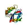
Mass: 18.015 Da / Num. of mol.: 103 / Source method: isolated from a natural source / Formula: H2O

-
Experimental details

-
Experiment

ExperimentMethod: X-RAY DIFFRACTION / Number of used crystals: 1

-
Sample preparation

CrystalDensity Matthews: 1.8 Å3/Da / Density % sol: 31.66 %
Crystal growTemperature: 298 K / Method: vapor diffusion, sitting drop / pH: 6.5
Details: 0.15 M Potassium thiocyanate, 0.1 M Sodium cacodylate pH 6.5, 20% v/v PEG 600

-
Data collection

DiffractionMean temperature: 100 K / Serial crystal experiment: N
Diffraction sourceSource: SYNCHROTRON / Site: SLS / Beamline: X06DA / Wavelength: 1.00001 Å
DetectorType: DECTRIS PILATUS 6M / Detector: PIXEL / Date: Jul 14, 2019
RadiationProtocol: SINGLE WAVELENGTH / Monochromatic (M) / Laue (L): M / Scattering type: x-ray
Radiation wavelengthWavelength: 1.00001 Å / Relative weight: 1
ReflectionResolution: 1.25→35.78 Å / Num. obs: 28291 / % possible obs: 98.5 % / Redundancy: 6.5 % / Biso Wilson estimate: 14.92 Å2 / CC1/2: 0.999 / Rmerge(I) obs: 0.046 / Rpim(I) all: 0.019 / Rrim(I) all: 0.05 / Net I/σ(I): 16.9 / Num. measured all: 184150 / Scaling rejects: 190
Reflection shell

Diffraction-ID: 1

Resolution (Å)Redundancy (%)Rmerge(I) obsNum. measured allNum. unique obsCC1/2Rpim(I) allRrim(I) allNet I/σ(I) obs% possible all
1.25-1.276.50.379950814690.9120.1610.4124.799.8
6.72-35.785.70.03810581860.9990.0170.04236.390.2

-
Phasing

PhasingMethod: molecular replacement
Phasing MRR rigid body: 0.578
Highest resolutionLowest resolution
Rotation35.78 Å4 Å

-
Processing

Software
NameVersionClassificationNB
PHENIX1.12_2829refinement
MOSFLMdata reduction
Aimlessdata scaling
MOLREP11.7.01phasing
PDB_EXTRACT3.25data extraction
RefinementMethod to determine structure: MOLECULAR REPLACEMENT
Starting model: Lpd1

Resolution: 1.25→28.158 Å / SU ML: 0.12 / Cross valid method: THROUGHOUT / σ(F): 1.4 / Phase error: 17.12 / Stereochemistry target values: ML
RfactorNum. reflection% reflectionSelection details
Rfree0.1879 1372 4.89 %RANDOM
Rwork0.1645 26701 --
obs0.1656 28073 98.38 %-
Solvent computationShrinkage radii: 0.9 Å / VDW probe radii: 1.11 Å / Solvent model: FLAT BULK SOLVENT MODEL
Displacement parametersBiso max: 60.92 Å2 / Biso mean: 22.6188 Å2 / Biso min: 9.55 Å2
Refinement stepCycle: final / Resolution: 1.25→28.158 Å
ProteinNucleic acidLigandSolventTotal
Num. atoms823 0 0 103 926
Biso mean---37.41 -
Num. residues----108
Refine LS restraints
Refine-IDTypeDev idealNumber
X-RAY DIFFRACTIONf_bond_d0.014851
X-RAY DIFFRACTIONf_angle_d1.4841150
X-RAY DIFFRACTIONf_chiral_restr0.156136
X-RAY DIFFRACTIONf_plane_restr0.009149
X-RAY DIFFRACTIONf_dihedral_angle_d8.477517
LS refinement shell

Refine-ID: X-RAY DIFFRACTION / Rfactor Rfree error: 0

Resolution (Å)Rfactor RfreeNum. reflection RfreeRfactor RworkNum. reflection Rwork% reflection obs (%)
1.25-1.29460.20761350.16162687100
1.2946-1.34640.17711500.1423265299
1.3464-1.40770.17771280.1324265898
1.4077-1.48190.18781270.1408254894
1.4819-1.57470.16151550.1252690100
1.5747-1.69630.19571530.13232688100
1.6963-1.86690.16761410.145267799
1.8669-2.1370.17751280.1414264097
2.137-2.69210.20961300.16882727100
2.6921-28.1580.18971250.1927273498

+
About Yorodumi

-
News

-
Feb 9, 2022. New format data for meta-information of EMDB entries

New format data for meta-information of EMDB entries

  • Version 3 of the EMDB header file is now the official format.
  • The previous official version 1.9 will be removed from the archive.

Related info.:EMDB header

External links:wwPDB to switch to version 3 of the EMDB data model

-
Aug 12, 2020. Covid-19 info

Covid-19 info

URL: https://pdbj.org/emnavi/covid19.php

New page: Covid-19 featured information page in EM Navigator.

Related info.:Covid-19 info / Mar 5, 2020. Novel coronavirus structure data

+
Mar 5, 2020. Novel coronavirus structure data

Novel coronavirus structure data

Related info.:Yorodumi Speices / Aug 12, 2020. Covid-19 info

External links:COVID-19 featured content - PDBj / Molecule of the Month (242):Coronavirus Proteases

+
Jan 31, 2019. EMDB accession codes are about to change! (news from PDBe EMDB page)

EMDB accession codes are about to change! (news from PDBe EMDB page)

  • The allocation of 4 digits for EMDB accession codes will soon come to an end. Whilst these codes will remain in use, new EMDB accession codes will include an additional digit and will expand incrementally as the available range of codes is exhausted. The current 4-digit format prefixed with “EMD-” (i.e. EMD-XXXX) will advance to a 5-digit format (i.e. EMD-XXXXX), and so on. It is currently estimated that the 4-digit codes will be depleted around Spring 2019, at which point the 5-digit format will come into force.
  • The EM Navigator/Yorodumi systems omit the EMD- prefix.

Related info.:Q: What is EMD? / ID/Accession-code notation in Yorodumi/EM Navigator

External links:EMDB Accession Codes are Changing Soon! / Contact to PDBj

+
Jul 12, 2017. Major update of PDB

Major update of PDB

  • wwPDB released updated PDB data conforming to the new PDBx/mmCIF dictionary.
  • This is a major update changing the version number from 4 to 5, and with Remediation, in which all the entries are updated.
  • In this update, many items about electron microscopy experimental information are reorganized (e.g. em_software).
  • Now, EM Navigator and Yorodumi are based on the updated data.

External links:wwPDB Remediation / Enriched Model Files Conforming to OneDep Data Standards Now Available in the PDB FTP Archive

-
Yorodumi

Thousand views of thousand structures

  • Yorodumi is a browser for structure data from EMDB, PDB, SASBDB, etc.
  • This page is also the successor to EM Navigator detail page, and also detail information page/front-end page for Omokage search.
  • The word "yorodu" (or yorozu) is an old Japanese word meaning "ten thousand". "mi" (miru) is to see.

Related info.:EMDB / PDB / SASBDB / Comparison of 3 databanks / Yorodumi Search / Aug 31, 2016. New EM Navigator & Yorodumi / Yorodumi Papers / Jmol/JSmol / Function and homology information / Changes in new EM Navigator and Yorodumi

Read more