 Movie
Movie Controller


+ Open data

- Basic information
| Entry | Database: PDB / ID: 6tm8 | ||||||
|---|---|---|---|---|---|---|---|
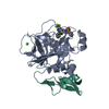
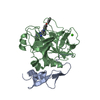
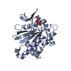
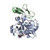
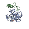
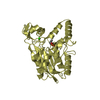
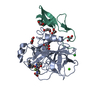
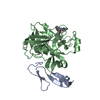
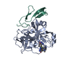
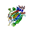
| Title | Crystal structure of glycoprotein D of Equine Herpesvirus Type 4 | ||||||
 Components Components | Envelope glycoprotein D | ||||||
 Keywords Keywords | VIRAL PROTEIN / glycoprotein D / equine herpesvirus type 4 / EHV-4 / viral entry | ||||||
| Function / homology | Herpesvirus glycoprotein D/GG/GX domain / Herpesvirus glycoprotein D/GG/GX domain / Immunoglobulin-like domain superfamily / viral envelope / membrane / Envelope glycoprotein D Function and homology information Function and homology information | ||||||
| Biological species |  Equid alphaherpesvirus 4 (Equine herpesvirus 4) Equid alphaherpesvirus 4 (Equine herpesvirus 4) | ||||||
| Method |  X-RAY DIFFRACTION / SYNCHROTRON / MOLECULAR REPLACEMENT / Resolution: 1.9 Å X-RAY DIFFRACTION / SYNCHROTRON / MOLECULAR REPLACEMENT / Resolution: 1.9 Å | ||||||
 Authors Authors | Kremling, V. / Loll, B. / Osterrieder, N. / Wahl, M. / Dahmani, I. / Chiantia, P. / Azab, W. | ||||||
| Funding support |  Germany, 1items Germany, 1items
| ||||||
 Citation Citation | Journal: Front Microbiol / Year: 2023 Title: Crystal structures of glycoprotein D of equine alphaherpesviruses reveal potential binding sites to the entry receptor MHC-I. Authors: Kremling, V. / Loll, B. / Pach, S. / Dahmani, I. / Weise, C. / Wolber, G. / Chiantia, S. / Wahl, M.C. / Osterrieder, N. / Azab, W. | ||||||
| History |
|
- Structure visualization
| Structure viewer | Molecule: MolmilJmol/JSmol |
|---|
- Downloads & links
-Download
| PDBx/mmCIF format | 6tm8.cif.gz | 148.9 KB | Display | PDBx/mmCIF format |
|---|---|---|---|---|
| PDB format | pdb6tm8.ent.gz | 95.2 KB | Display | PDB format |
| PDBx/mmJSON format | 6tm8.json.gz | Tree view | PDBx/mmJSON format | |
| Others |  Other downloads Other downloads |
-Validation report
| Arichive directory | https://data.pdbj.org/pub/pdb/validation_reports/tm/6tm8ftp://data.pdbj.org/pub/pdb/validation_reports/tm/6tm8 | HTTPS FTP |
|---|
-Related structure data
| Related structure data | 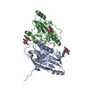 6sqjSC S: Starting model for refinement C: citing same article ( |
|---|---|
| Similar structure data |
-Links
 PDBj
PDBj- Assembly
| Deposited unit | 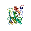
| ||||||||||||
|---|---|---|---|---|---|---|---|---|---|---|---|---|---|
| 1 |
| ||||||||||||
| Unit cell |
|
-Components
| #1: Protein | Mass: 38023.652 Da / Num. of mol.: 1 Source method: isolated from a genetically manipulated source Source: (gene. exp.) Equid alphaherpesvirus 4 (Equine herpesvirus 4)Gene: ORF72 / Plasmid: pMultiBac / Cell line (production host): H5 / Production host:  Trichoplusia ni (cabbage looper) / References: UniProt: A0A0Y0A4Z5 Trichoplusia ni (cabbage looper) / References: UniProt: A0A0Y0A4Z5 | ||||||
|---|---|---|---|---|---|---|---|
| #2: Chemical | ChemComp-GOL /   Mass: 92.094 Da / Num. of mol.: 4 / Source method: obtained synthetically / Formula: C3H8O3 / Feature type: SUBJECT OF INVESTIGATION Mass: 92.094 Da / Num. of mol.: 4 / Source method: obtained synthetically / Formula: C3H8O3 / Feature type: SUBJECT OF INVESTIGATION#3: Water | ChemComp-HOH / |  Mass: 18.015 Da / Num. of mol.: 174 / Source method: isolated from a natural source / Formula: H2O Mass: 18.015 Da / Num. of mol.: 174 / Source method: isolated from a natural source / Formula: H2OHas ligand of interest | Y | Has protein modification | Y | |
-Experimental details
-Experiment
| Experiment | Method: X-RAY DIFFRACTION / Number of used crystals: 1 |
|---|
- Sample preparation
| Crystal | Density Matthews: 1.99 Å3/Da / Density % sol: 38.32 % |
|---|---|
| Crystal grow | Temperature: 291 K / Method: vapor diffusion, sitting drop / Details: 100 mM MgCl2, 100 mM MES pH 6.5, 30% (w/v) PEG 400 |
-Data collection
| Diffraction | Mean temperature: 100 K / Serial crystal experiment: N |
|---|---|
| Diffraction source | Source: SYNCHROTRON / Site: BESSY / Beamline: 14.1 / Wavelength: 0.91841 Å |
| Detector | Type: DECTRIS PILATUS 6M / Detector: PIXEL / Date: Nov 6, 2019 |
| Radiation | Protocol: SINGLE WAVELENGTH / Monochromatic (M) / Laue (L): M / Scattering type: x-ray |
| Radiation wavelength | Wavelength: 0.91841 Å / Relative weight: 1 |
| Reflection | Resolution: 1.9→50 Å / Num. obs: 23671 / % possible obs: 99.5 % / Redundancy: 5.9 % / Biso Wilson estimate: 32 Å2 / CC1/2: 0.995 / Rrim(I) all: 0.174 / Net I/σ(I): 8.96 |
| Reflection shell | Resolution: 1.9→2.01 Å / Redundancy: 3.5 % / Num. unique obs: 10835 / CC1/2: 0.409 / Rrim(I) all: 1.268 / % possible all: 78.2 |
- Processing
| Software |
| |||||||||||||||||||||||||||||||||||||||||||||||||||||||||||||||
|---|---|---|---|---|---|---|---|---|---|---|---|---|---|---|---|---|---|---|---|---|---|---|---|---|---|---|---|---|---|---|---|---|---|---|---|---|---|---|---|---|---|---|---|---|---|---|---|---|---|---|---|---|---|---|---|---|---|---|---|---|---|---|---|---|
| Refinement | Method to determine structure: MOLECULAR REPLACEMENT Starting model: 6SQJ Resolution: 1.9→46.17 Å / SU ML: 0.2323 / Cross valid method: FREE R-VALUE / σ(F): 1.36 / Phase error: 20.9286
| |||||||||||||||||||||||||||||||||||||||||||||||||||||||||||||||
| Solvent computation | Shrinkage radii: 0.9 Å / VDW probe radii: 1.11 Å | |||||||||||||||||||||||||||||||||||||||||||||||||||||||||||||||
| Displacement parameters | Biso mean: 31.08 Å2 | |||||||||||||||||||||||||||||||||||||||||||||||||||||||||||||||
| Refinement step | Cycle: LAST / Resolution: 1.9→46.17 Å
| |||||||||||||||||||||||||||||||||||||||||||||||||||||||||||||||
| Refine LS restraints |
| |||||||||||||||||||||||||||||||||||||||||||||||||||||||||||||||
| LS refinement shell |
| |||||||||||||||||||||||||||||||||||||||||||||||||||||||||||||||
| Refinement TLS params. | Method: refined / Origin x: -18.3823856533 Å / Origin y: -2.65350132849 Å / Origin z: 8.15552908134 Å
| |||||||||||||||||||||||||||||||||||||||||||||||||||||||||||||||
| Refinement TLS group | Selection details: all |
