 Movie
Movie Controller

[English] 日本語
 Yorodumi
- PDB-6tku: Crystal structure of a capsule-specific depolymerase produced by ... -
+ Open data

- Basic information
| Entry | Database: PDB / ID: 6tku | ||||||
|---|---|---|---|---|---|---|---|
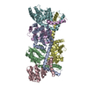
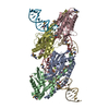
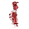
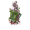
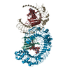
| Title | Crystal structure of a capsule-specific depolymerase produced by Klebsiella phage | ||||||
 Components Components | depolymerase KP32gp38 | ||||||
 Keywords Keywords | HYDROLASE / Klebsiella pneumoniae capsule / phage depolymerase / tail fiber branching system | ||||||
| Function / homology | symbiont entry into host cell via disruption of host cell glycocalyx / symbiont entry into host cell via disruption of host cell envelope / virus tail / adhesion receptor-mediated virion attachment to host cell / Pectin lyase fold / Pectin lyase fold/virulence factor / Depolymerase 2, capsule K21-specific Function and homology information Function and homology information | ||||||
| Biological species |  Klebsiella phage KP32 (virus) Klebsiella phage KP32 (virus) | ||||||
| Method |  X-RAY DIFFRACTION / SYNCHROTRON / SAD / Resolution: 1.8 Å X-RAY DIFFRACTION / SYNCHROTRON / SAD / Resolution: 1.8 Å | ||||||
 Authors Authors | Berisio, R. / Squeglia, F. | ||||||
| Funding support |  Italy, 1items Italy, 1items
| ||||||
 Citation Citation | Journal: Structure / Year: 2020 Title: Structural and Functional Studies of a Klebsiella Phage Capsule Depolymerase Tailspike: Mechanistic Insights into Capsular Degradation. Authors: Squeglia, F. / Maciejewska, B. / Latka, A. / Ruggiero, A. / Briers, Y. / Drulis-Kawa, Z. / Berisio, R. | ||||||
| History |
|
- Structure visualization
| Structure viewer | Molecule: MolmilJmol/JSmol |
|---|
- Downloads & links
-Download
| PDBx/mmCIF format | 6tku.cif.gz | 129.3 KB | Display | PDBx/mmCIF format |
|---|---|---|---|---|
| PDB format | pdb6tku.ent.gz | 97.5 KB | Display | PDB format |
| PDBx/mmJSON format | 6tku.json.gz | Tree view | PDBx/mmJSON format | |
| Others |  Other downloads Other downloads |
-Validation report
| Arichive directory | https://data.pdbj.org/pub/pdb/validation_reports/tk/6tkuftp://data.pdbj.org/pub/pdb/validation_reports/tk/6tku | HTTPS FTP |
|---|
-Related structure data
| Similar structure data |
|---|


-Links
 PDBj
PDBj- Assembly
| Deposited unit | 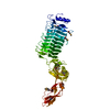
| |||||||||||||||||||||
|---|---|---|---|---|---|---|---|---|---|---|---|---|---|---|---|---|---|---|---|---|---|---|
| 1 | 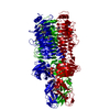
| |||||||||||||||||||||
| Unit cell |
| |||||||||||||||||||||
| Components on special symmetry positions |
|
-Components
| #1: Protein | Mass: 63317.254 Da / Num. of mol.: 1 Source method: isolated from a genetically manipulated source Source: (gene. exp.) Klebsiella phage KP32 (virus) / Gene: 38 / Production host:  |
|---|---|
| #2: Water | ChemComp-HOH /  Mass: 18.015 Da / Num. of mol.: 444 / Source method: isolated from a natural source / Formula: H2O Mass: 18.015 Da / Num. of mol.: 444 / Source method: isolated from a natural source / Formula: H2O |
| Has ligand of interest | N |
| Has protein modification | Y |
-Experimental details
-Experiment
| Experiment | Method: X-RAY DIFFRACTION / Number of used crystals: 1 |
|---|
- Sample preparation
| Crystal | Density Matthews: 2.46 Å3/Da / Density % sol: 50.07 % |
|---|---|
| Crystal grow | Temperature: 293 K / Method: vapor diffusion, hanging drop Details: 0.2 M Ammonium citrate tribasic pH 7.0 and 20% w/v polyethylene glycol 3,350. |
-Data collection
| Diffraction | Mean temperature: 100 K / Serial crystal experiment: N |
|---|---|
| Diffraction source | Source: SYNCHROTRON / Site: ELETTRA / Beamline: 5.2R / Wavelength: 0.9759 Å |
| Detector | Type: DECTRIS PILATUS 6M / Detector: PIXEL / Date: May 7, 2019 |
| Radiation | Protocol: SINGLE WAVELENGTH / Monochromatic (M) / Laue (L): M / Scattering type: x-ray |
| Radiation wavelength | Wavelength: 0.9759 Å / Relative weight: 1 |
| Reflection | Resolution: 1.8→30 Å / Num. obs: 59968 / % possible obs: 99.9 % / Redundancy: 13.6 % / Rmerge(I) obs: 0.05 / Net I/σ(I): 19.7 |
| Reflection shell | Resolution: 1.8→1.84 Å / Rmerge(I) obs: 0.232 / Num. unique obs: 4226 |
- Processing
| Software |
| ||||||||||||||||||||||||||||||||||||||||||||||||||||||||||||
|---|---|---|---|---|---|---|---|---|---|---|---|---|---|---|---|---|---|---|---|---|---|---|---|---|---|---|---|---|---|---|---|---|---|---|---|---|---|---|---|---|---|---|---|---|---|---|---|---|---|---|---|---|---|---|---|---|---|---|---|---|---|
| Refinement | Method to determine structure: SAD / Resolution: 1.8→14.91 Å / Cor.coef. Fo:Fc: 0.959 / Cor.coef. Fo:Fc free: 0.933 / SU B: 3.575 / SU ML: 0.107 / Cross valid method: THROUGHOUT / σ(F): 0 / ESU R: 0.132 / ESU R Free: 0.132 Details: HYDROGENS HAVE BEEN ADDED IN THE RIDING POSITIONS U VALUES
| ||||||||||||||||||||||||||||||||||||||||||||||||||||||||||||
| Solvent computation | Ion probe radii: 0.8 Å / Shrinkage radii: 0.8 Å / VDW probe radii: 1.2 Å | ||||||||||||||||||||||||||||||||||||||||||||||||||||||||||||
| Displacement parameters | Biso max: 102.26 Å2 / Biso mean: 40.1911 Å2 / Biso min: 12.16 Å2
| ||||||||||||||||||||||||||||||||||||||||||||||||||||||||||||
| Refinement step | Cycle: final / Resolution: 1.8→14.91 Å
| ||||||||||||||||||||||||||||||||||||||||||||||||||||||||||||
| Refine LS restraints |
| ||||||||||||||||||||||||||||||||||||||||||||||||||||||||||||
| LS refinement shell | Resolution: 1.8→1.846 Å / Rfactor Rfree error: 0 / Total num. of bins used: 20
|
