Mass: 251.242 Da / Num. of mol.: 4 / Source method: obtained synthetically / Formula: C10H13N5O3
Sequence details
THE STRUCTURE DOES NOT CONTAIN RESIDUES GLY220 TO ASP222 OF ORAE PRESENT IN THE UNP SEQUENCE. WE DO ...THE STRUCTURE DOES NOT CONTAIN RESIDUES GLY220 TO ASP222 OF ORAE PRESENT IN THE UNP SEQUENCE. WE DO NOT FIND ANY EVIDENCE FOR THESE RESIDUES FROM CLOSTRIDIUM STICKLANDII GENOMIC DNA SEQUENCING OR BY ALIGNMENT BETWEEN THE ORAE PROTEIN SEQUENCES FROM CLOSTRIDIUM STICKLANDII AND CLOSTRIDIUM DIFFICILE (ACCESSION NUMBER ZP_05349629; 79% SEQUENCE IDENTITY) WHICH ALSO SHOWS THAT THE CORRESPONDING ORAE CODING SEQUENCE FROM THE LATTER ORGANISM ALSO DOES NOT CONTAIN THE THREE AMINO ACID GID INSERT (A SIMILAR CONCLUSION CAN BE REACHED BY ALIGNMENT WITH VARIOUS THERMOANAEROBACTER SP)
-
Experimental details
-
Experiment
Experiment
Method: X-RAY DIFFRACTION / Number of used crystals: 1
-
Sample preparation
Crystal
Density Matthews: 2.36 Å3/Da / Density % sol: 47.96 %
Crystal grow
Temperature: 294 K / Method: vapor diffusion, sitting drop / pH: 8 Details: A protein solution (8 mg/ml 4,5-OAM, 5 mM 2-mercaptoethanol, 10 mM Tris HCl pH 8.0, 2 mM PLP, and 2 mM AdoCbl) was mixed in a 1:1 ratio with precipitant solution [(0.1 M Tris HCl, pH 8.0, 0. ...Details: A protein solution (8 mg/ml 4,5-OAM, 5 mM 2-mercaptoethanol, 10 mM Tris HCl pH 8.0, 2 mM PLP, and 2 mM AdoCbl) was mixed in a 1:1 ratio with precipitant solution [(0.1 M Tris HCl, pH 8.0, 0.2 M MgCl2, 25 % (wt/vol) polyethylene glycol 2000 mono-methylether] under red light at room temperature, VAPOR DIFFUSION, SITTING DROP, temperature 294K
In the structure databanks used in Yorodumi, some data are registered as the other names, "COVID-19 virus" and "2019-nCoV". Here are the details of the virus and the list of structure data.
Jan 31, 2019. EMDB accession codes are about to change! (news from PDBe EMDB page)
EMDB accession codes are about to change! (news from PDBe EMDB page)
The allocation of 4 digits for EMDB accession codes will soon come to an end. Whilst these codes will remain in use, new EMDB accession codes will include an additional digit and will expand incrementally as the available range of codes is exhausted. The current 4-digit format prefixed with “EMD-” (i.e. EMD-XXXX) will advance to a 5-digit format (i.e. EMD-XXXXX), and so on. It is currently estimated that the 4-digit codes will be depleted around Spring 2019, at which point the 5-digit format will come into force.
The EM Navigator/Yorodumi systems omit the EMD- prefix.
Related info.:Q: What is EMD? / ID/Accession-code notation in Yorodumi/EM Navigator
Yorodumi is a browser for structure data from EMDB, PDB, SASBDB, etc.
This page is also the successor to EM Navigator detail page, and also detail information page/front-end page for Omokage search.
The word "yorodu" (or yorozu) is an old Japanese word meaning "ten thousand". "mi" (miru) is to see.
Related info.:EMDB / PDB / SASBDB / Comparison of 3 databanks / Yorodumi Search / Aug 31, 2016. New EM Navigator & Yorodumi / Yorodumi Papers / Jmol/JSmol / Function and homology information / Changes in new EM Navigator and Yorodumi
 Movie
Movie Controller

 Yorodumi
 Open data
 Basic information
 Components
 Keywords
 Function and homology information
 Clostridium sticklandii (bacteria)
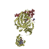
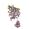
 X-RAY DIFFRACTION /
  Authors
 Citation
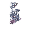
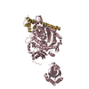
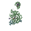
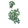
 Structure visualization
 Downloads & links
 Other downloads


 PDBj
PDBj Assembly
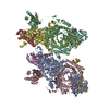


 Mass: 1330.356 Da / Num. of mol.: 4 / Source method: obtained synthetically / Formula: C62H89CoN13O14P
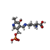
 Mass: 361.288 Da / Num. of mol.: 4 / Source method: obtained synthetically / Formula: C13H20N3O7P
 Mass: 251.242 Da / Num. of mol.: 4 / Source method: obtained synthetically / Formula: C10H13N5O3
 Sample preparation
 / Beamline: I02 / Wavelength: 1.072 Å
 Processing