 Movie
Movie Controller

[English] 日本語
 Yorodumi
- PDB-6tk1: Femtosecond to millisecond structural changes in a light-driven s... -
+ Open data

- Basic information
| Entry | Database: PDB / ID: 6tk1 | ||||||||||||||||||||||||||||||
|---|---|---|---|---|---|---|---|---|---|---|---|---|---|---|---|---|---|---|---|---|---|---|---|---|---|---|---|---|---|---|---|
| Title | Femtosecond to millisecond structural changes in a light-driven sodium pump: 20ms structure of KR2 with extrapolated, light and dark datasets | ||||||||||||||||||||||||||||||
 Components Components | Sodium pumping rhodopsin | ||||||||||||||||||||||||||||||
 Keywords Keywords | MEMBRANE PROTEIN / Sodium pumping rhodopsin / time-resolved / serial femtosecond crystallograpy / room-temperature | ||||||||||||||||||||||||||||||
| Function / homology |  Function and homology information Function and homology information | ||||||||||||||||||||||||||||||
| Biological species |  Dokdonia eikasta (bacteria) Dokdonia eikasta (bacteria) | ||||||||||||||||||||||||||||||
| Method |  X-RAY DIFFRACTION / FREE ELECTRON LASER / MOLECULAR REPLACEMENT / Resolution: 2.5 Å X-RAY DIFFRACTION / FREE ELECTRON LASER / MOLECULAR REPLACEMENT / Resolution: 2.5 Å | ||||||||||||||||||||||||||||||
 Authors Authors | Skopintsev, P. / Ehrenberg, D. / Weinert, T. / James, D. / Kar, R. / Johnson, P. / Ozerov, D. / Furrer, A. / Martiel, I. / Dworkowski, F. ...Skopintsev, P. / Ehrenberg, D. / Weinert, T. / James, D. / Kar, R. / Johnson, P. / Ozerov, D. / Furrer, A. / Martiel, I. / Dworkowski, F. / Nass, K. / Knopp, G. / Cirelli, C. / Gashi, D. / Mous, S. / Wranik, M. / Gruhl, T. / Kekilli, D. / Bruenle, S. / Deupi, X. / Schertler, G.F.X. / Benoit, R. / Panneels, V. / Nogly, P. / Schapiro, I. / Milne, C. / Heberle, J. / Standfuss, J. | ||||||||||||||||||||||||||||||
| Funding support |  Switzerland, Switzerland,  Germany, Germany,  Israel, 9items Israel, 9items
| ||||||||||||||||||||||||||||||
 Citation Citation | Journal: Nature / Year: 2020 Title: Femtosecond-to-millisecond structural changes in a light-driven sodium pump. Authors: Skopintsev, P. / Ehrenberg, D. / Weinert, T. / James, D. / Kar, R.K. / Johnson, P.J.M. / Ozerov, D. / Furrer, A. / Martiel, I. / Dworkowski, F. / Nass, K. / Knopp, G. / Cirelli, C. / Arrell, ...Authors: Skopintsev, P. / Ehrenberg, D. / Weinert, T. / James, D. / Kar, R.K. / Johnson, P.J.M. / Ozerov, D. / Furrer, A. / Martiel, I. / Dworkowski, F. / Nass, K. / Knopp, G. / Cirelli, C. / Arrell, C. / Gashi, D. / Mous, S. / Wranik, M. / Gruhl, T. / Kekilli, D. / Brunle, S. / Deupi, X. / Schertler, G.F.X. / Benoit, R.M. / Panneels, V. / Nogly, P. / Schapiro, I. / Milne, C. / Heberle, J. / Standfuss, J. | ||||||||||||||||||||||||||||||
| History |
|
- Structure visualization
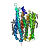
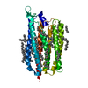
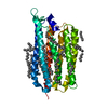
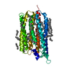
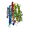
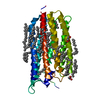
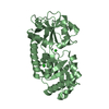
| Structure viewer | Molecule: MolmilJmol/JSmol |
|---|
- Downloads & links
-Download
| PDBx/mmCIF format | 6tk1.cif.gz | 79.6 KB | Display | PDBx/mmCIF format |
|---|---|---|---|---|
| PDB format | pdb6tk1.ent.gz | 55.9 KB | Display | PDB format |
| PDBx/mmJSON format | 6tk1.json.gz | Tree view | PDBx/mmJSON format | |
| Others |  Other downloads Other downloads |
-Validation report
| Arichive directory | https://data.pdbj.org/pub/pdb/validation_reports/tk/6tk1ftp://data.pdbj.org/pub/pdb/validation_reports/tk/6tk1 | HTTPS FTP |
|---|
-Related structure data
| Related structure data |  6tk2C  6tk3C  6tk4C  6tk5C  6tk6C  6tk7C  3x3cS S: Starting model for refinement C: citing same article ( |
|---|---|
| Similar structure data |

-Links
 PDBj
PDBj


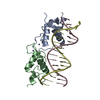
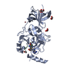
- Assembly
| Deposited unit | 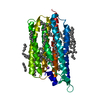
| ||||||||
|---|---|---|---|---|---|---|---|---|---|
| 1 |
| ||||||||
| Unit cell |
| ||||||||
| Components on special symmetry positions |
|
-Components
| #1: Protein | Mass: 32894.859 Da / Num. of mol.: 1 Source method: isolated from a genetically manipulated source Source: (gene. exp.) Dokdonia eikasta (bacteria) / Gene: NaR / Production host: | ||||||||
|---|---|---|---|---|---|---|---|---|---|
| #2: Chemical | ChemComp-RET /   Mass: 284.436 Da / Num. of mol.: 1 / Source method: obtained synthetically / Formula: C20H28O / Feature type: SUBJECT OF INVESTIGATION Mass: 284.436 Da / Num. of mol.: 1 / Source method: obtained synthetically / Formula: C20H28O / Feature type: SUBJECT OF INVESTIGATION | ||||||||
| #3: Chemical | ChemComp-LFA /   Mass: 282.547 Da / Num. of mol.: 26 / Source method: obtained synthetically / Formula: C20H42 Mass: 282.547 Da / Num. of mol.: 26 / Source method: obtained synthetically / Formula: C20H42#4: Chemical | ChemComp-NA / |   Mass: 22.990 Da / Num. of mol.: 1 / Source method: obtained synthetically / Formula: Na / Feature type: SUBJECT OF INVESTIGATION Mass: 22.990 Da / Num. of mol.: 1 / Source method: obtained synthetically / Formula: Na / Feature type: SUBJECT OF INVESTIGATION#5: Water | ChemComp-HOH / |  Mass: 18.015 Da / Num. of mol.: 47 / Source method: isolated from a natural source / Formula: H2O Mass: 18.015 Da / Num. of mol.: 47 / Source method: isolated from a natural source / Formula: H2OHas ligand of interest | Y | Has protein modification | Y | |
-Experimental details
-Experiment
| Experiment | Method: X-RAY DIFFRACTION / Number of used crystals: 1 |
|---|
- Sample preparation
| Crystal | Density Matthews: 3.14 Å3/Da / Density % sol: 60.84 % |
|---|---|
| Crystal grow | Temperature: 291 K / Method: lipidic cubic phase / pH: 4.4 Details: 200 mM Sodium Acetate pH 4.4, 150 mM MgCl2, 35% PEG 200 |
-Data collection
| Diffraction | Mean temperature: 293 K / Serial crystal experiment: Y |
|---|---|
| Diffraction source | Source: FREE ELECTRON LASER / Site: SwissFEL ARAMIS / Beamline: ESA / Wavelength: 1 Å |
| Detector | Type: PSI JUNGFRAU 16M / Detector: PIXEL / Date: Feb 25, 2019 |
| Radiation | Protocol: SINGLE WAVELENGTH / Monochromatic (M) / Laue (L): M / Scattering type: x-ray |
| Radiation wavelength | Wavelength: 1 Å / Relative weight: 1 |
| Reflection | Resolution: 2.5→12.4 Å / Num. obs: 13268 / % possible obs: 89.9 % / Redundancy: 1 % / CC1/2: 1 / Net I/σ(I): 1 |
| Reflection shell | Resolution: 2.5→2.66 Å / Num. unique obs: 1871 / CC1/2: 1 / % possible all: 78 |
| Serial crystallography measurement | Focal spot size: 3.5 µm2 / XFEL pulse repetition rate: 50 Hz |
| Serial crystallography sample delivery | Description: High viscosity injector / Method: injection |
| Serial crystallography sample delivery injection | Carrier solvent: LCP / Jet diameter: 75 µm / Power by: HPLC |
- Processing
| Software |
| ||||||||||||||||||||||||||||||||||||||||||
|---|---|---|---|---|---|---|---|---|---|---|---|---|---|---|---|---|---|---|---|---|---|---|---|---|---|---|---|---|---|---|---|---|---|---|---|---|---|---|---|---|---|---|---|
| Refinement | Method to determine structure: MOLECULAR REPLACEMENT Starting model: 3x3c Resolution: 2.5→12.286 Å / SU ML: 0.55 / Cross valid method: THROUGHOUT / σ(F): 0.01 / Phase error: 40.23
| ||||||||||||||||||||||||||||||||||||||||||
| Solvent computation | Shrinkage radii: 0.9 Å / VDW probe radii: 1.11 Å | ||||||||||||||||||||||||||||||||||||||||||
| Displacement parameters | Biso max: 113.13 Å2 / Biso mean: 55.0597 Å2 / Biso min: 27.74 Å2 | ||||||||||||||||||||||||||||||||||||||||||
| Refinement step | Cycle: final / Resolution: 2.5→12.286 Å
| ||||||||||||||||||||||||||||||||||||||||||
| LS refinement shell | Refine-ID: X-RAY DIFFRACTION / Rfactor Rfree error: 0
|
