[English] 日本語
Yorodumi
- PDB-6thk: Structural mechanism of pyocin S5 import into Pseudomonas aeruginosa -

+
Open data


ID or keywords:

Loading...

-
Basic information

Entry
Database: PDB / ID: 6thk
TitleStructural mechanism of pyocin S5 import into Pseudomonas aeruginosa
ComponentsPyocin S5
KeywordsANTIMICROBIAL PROTEIN / Bacteriocin / Pyocin / Antibiotic / Ionophore
Function / homology
Function and homology information


pore-forming activity / defense response to Gram-negative bacterium / killing of cells of another organism / metal ion binding / membrane
Similarity search - Function
Channel forming colicin, C-terminal cytotoxic / Channel forming colicin, C-terminal domain superfamily / Colicin pore forming domain / Channel forming colicins signature.
Similarity search - Domain/homology
Biological speciesPseudomonas aeruginosa PAO1 (bacteria)
MethodX-RAY DIFFRACTION / SYNCHROTRON / SAD / Resolution: 2.2 Å
AuthorsBehrens, H.M. / Kleanthous, C. / Lowe, E.D.
Funding support United Kingdom, 2items
OrganizationGrant numberCountry
Wellcome TrustInfection, Immunology and Translational Medicine studentship United Kingdom
Wellcome Trust201505/Z/16/Z United Kingdom
Citation
Journal: Mbio / Year: 2020
Title: Pyocin S5 Import into Pseudomonas aeruginosa Reveals a Generic Mode of Bacteriocin Transport.
Authors: Behrens, H.M. / Lowe, E.D. / Gault, J. / Housden, N.G. / Kaminska, R. / Weber, T.M. / Thompson, C.M.A. / Mislin, G.L.A. / Schalk, I.J. / Walker, D. / Robinson, C.V. / Kleanthous, C.
#1: Journal: Biorxiv / Year: 2019
Title: Pyocin S5 import into Pseudomonas aeruginosa reveals a generic mode of bacteriocin transport
Authors: Behrens, H.M. / Lowe, E.D. / Gault, J. / Housden, N.G. / Kaminska, R. / Weber, T.M. / Thompson, C. / Mislin, G.L.A. / Schalk, I.J. / Walker, D. / Robinson, C.V. / Kleanthous, C.
History
DepositionNov 20, 2019Deposition site: PDBE / Processing site: PDBE
Revision 1.0Mar 4, 2020Provider: repository / Type: Initial release
Revision 1.1Mar 25, 2020Group: Database references / Category: citation / citation_author
Revision 1.2May 15, 2024Group: Data collection / Database references / Derived calculations
Category: chem_comp_atom / chem_comp_bond ...chem_comp_atom / chem_comp_bond / citation / database_2 / pdbx_struct_conn_angle / struct_conn
Item: _citation.journal_id_ISSN / _database_2.pdbx_DOI ..._citation.journal_id_ISSN / _database_2.pdbx_DOI / _database_2.pdbx_database_accession / _pdbx_struct_conn_angle.ptnr1_auth_comp_id / _pdbx_struct_conn_angle.ptnr1_auth_seq_id / _pdbx_struct_conn_angle.ptnr1_label_asym_id / _pdbx_struct_conn_angle.ptnr1_label_atom_id / _pdbx_struct_conn_angle.ptnr1_label_comp_id / _pdbx_struct_conn_angle.ptnr1_label_seq_id / _pdbx_struct_conn_angle.ptnr2_auth_seq_id / _pdbx_struct_conn_angle.ptnr2_label_asym_id / _pdbx_struct_conn_angle.ptnr2_symmetry / _pdbx_struct_conn_angle.ptnr3_auth_comp_id / _pdbx_struct_conn_angle.ptnr3_auth_seq_id / _pdbx_struct_conn_angle.ptnr3_label_asym_id / _pdbx_struct_conn_angle.ptnr3_label_atom_id / _pdbx_struct_conn_angle.ptnr3_label_comp_id / _pdbx_struct_conn_angle.ptnr3_label_seq_id / _pdbx_struct_conn_angle.ptnr3_symmetry / _pdbx_struct_conn_angle.value / _struct_conn.pdbx_dist_value / _struct_conn.ptnr1_auth_comp_id / _struct_conn.ptnr1_auth_seq_id / _struct_conn.ptnr1_label_asym_id / _struct_conn.ptnr1_label_atom_id / _struct_conn.ptnr1_label_comp_id / _struct_conn.ptnr1_label_seq_id / _struct_conn.ptnr2_auth_comp_id / _struct_conn.ptnr2_auth_seq_id / _struct_conn.ptnr2_label_asym_id / _struct_conn.ptnr2_label_atom_id / _struct_conn.ptnr2_label_comp_id / _struct_conn.ptnr2_symmetry

-
Structure visualization

Structure viewerMolecule:
MolmilJmol/JSmol

Downloads & links

-
Assembly

Deposited unit
A: Pyocin S5
hetero molecules


Theoretical massNumber of molelcules
Total (without water)57,83810
Polymers57,2231
Non-polymers6159
Water1,51384
1


  • Idetical with deposited unit
  • defined by author&software
TypeNameSymmetry operationNumber
identity operation1_555x,y,z1
Buried area870 Å2
ΔGint-184 kcal/mol
Surface area26890 Å2
MethodPISA
Unit cell
Length a, b, c (Å)50.240, 53.870, 104.800
Angle α, β, γ (deg.)90.000, 95.150, 90.000
Int Tables number4
Space group name H-MP1211

-
Components

#1: Protein Pyocin S5


Mass: 57223.105 Da / Num. of mol.: 1 / Mutation: E215Q
Source method: isolated from a genetically manipulated source
Source: (gene. exp.) Pseudomonas aeruginosa PAO1 (bacteria) / Gene: pyoS5, PA0985 / Production host: Escherichia coli BL21(DE3) (bacteria) / References: UniProt: Q9I4Y4
#2: Chemical ChemComp-GOL / GLYCEROL / GLYCERIN / PROPANE-1,2,3-TRIOL


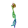
Mass: 92.094 Da / Num. of mol.: 1 / Source method: obtained synthetically / Formula: C3H8O3
#3: Chemical
ChemComp-ZN / ZINC ION


Mass: 65.409 Da / Num. of mol.: 8 / Source method: obtained synthetically / Formula: Zn
#4: Water ChemComp-HOH / water


Mass: 18.015 Da / Num. of mol.: 84 / Source method: isolated from a natural source / Formula: H2O
Has ligand of interestN

-
Experimental details

-
Experiment

ExperimentMethod: X-RAY DIFFRACTION / Number of used crystals: 1

-
Sample preparation

CrystalDensity Matthews: 2.47 Å3/Da / Density % sol: 50.17 %
Crystal growTemperature: 291.15 K / Method: vapor diffusion, sitting drop
Details: 10% w/v PEG 3000, 0.1 M sodium acetate, 0.1 M zinc acetate, pH 4.5 protein buffer: 25 mL Tris-HCl pH 7.5, 150 mM NaCl

-
Data collection

DiffractionMean temperature: 100 K / Serial crystal experiment: N
Diffraction sourceSource: SYNCHROTRON / Site: ESRF / Beamline: MASSIF-3 / Wavelength: 0.9679 Å
DetectorType: DECTRIS EIGER X 4M / Detector: PIXEL / Date: May 6, 2016
RadiationProtocol: SINGLE WAVELENGTH / Monochromatic (M) / Laue (L): M / Scattering type: x-ray
Radiation wavelengthWavelength: 0.9679 Å / Relative weight: 1
ReflectionResolution: 2.2→53.7 Å / Num. obs: 28285 / % possible obs: 98.8 % / Redundancy: 4.4 % / Biso Wilson estimate: 41.76 Å2 / CC1/2: 0.996 / Rmerge(I) obs: 0.138 / Rpim(I) all: 0.113 / Rrim(I) all: 0.179 / Χ2: 1.01 / Net I/σ(I): 4.9
Reflection shellResolution: 2.2→2.27 Å / Redundancy: 4.6 % / Rmerge(I) obs: 1.946 / Mean I/σ(I) obs: 0.7 / Num. unique obs: 2467 / CC1/2: 0.728 / Rpim(I) all: 1.594 / Rrim(I) all: 2.52 / Χ2: 1.11 / % possible all: 99.7

-
Processing

Software
NameVersionClassification
BUSTER2.10.3refinement
DIALSdata reduction
Aimlessdata scaling
PHASERphasing
RefinementMethod to determine structure: SAD / Resolution: 2.2→52.19 Å / Cor.coef. Fo:Fc: 0.897 / Cor.coef. Fo:Fc free: 0.856 / SU R Cruickshank DPI: 0.265 / Cross valid method: THROUGHOUT / σ(F): 0 / SU R Blow DPI: 0.256 / SU Rfree Blow DPI: 0.216 / SU Rfree Cruickshank DPI: 0.221
RfactorNum. reflection% reflectionSelection details
Rfree0.275 1301 4.62 %RANDOM
Rwork0.225 ---
obs0.227 28161 98.3 %-
Displacement parametersBiso max: 134.41 Å2 / Biso mean: 63.91 Å2 / Biso min: 30 Å2
Baniso -1Baniso -2Baniso -3
1-22.1836 Å20 Å214.0936 Å2
2--2.9421 Å20 Å2
3----25.1257 Å2
Refinement stepCycle: final / Resolution: 2.2→52.19 Å
ProteinNucleic acidLigandSolventTotal
Num. atoms3633 0 14 84 3731
Biso mean--66.87 60.53 -
Num. residues----466
Refine LS restraints
Refine-IDTypeNumberRestraint functionWeightDev ideal
X-RAY DIFFRACTIONt_dihedral_angle_d1335SINUSOIDAL2
X-RAY DIFFRACTIONt_trig_c_planes104HARMONIC2
X-RAY DIFFRACTIONt_gen_planes534HARMONIC5
X-RAY DIFFRACTIONt_it3712HARMONIC20
X-RAY DIFFRACTIONt_nbd1SEMIHARMONIC5
X-RAY DIFFRACTIONt_improper_torsion
X-RAY DIFFRACTIONt_pseud_angle
X-RAY DIFFRACTIONt_chiral_improper_torsion499SEMIHARMONIC5
X-RAY DIFFRACTIONt_sum_occupancies
X-RAY DIFFRACTIONt_utility_distance
X-RAY DIFFRACTIONt_utility_angle
X-RAY DIFFRACTIONt_utility_torsion
X-RAY DIFFRACTIONt_ideal_dist_contact4362SEMIHARMONIC4
X-RAY DIFFRACTIONt_bond_d3712HARMONIC20.01
X-RAY DIFFRACTIONt_angle_deg5014HARMONIC21.08
X-RAY DIFFRACTIONt_omega_torsion2.51
X-RAY DIFFRACTIONt_other_torsion21
LS refinement shellResolution: 2.2→2.28 Å / Rfactor Rfree error: 0 / Total num. of bins used: 14
RfactorNum. reflection% reflection
Rfree0.3093 138 4.62 %
Rwork0.2573 2848 -
all0.26 2986 -
obs--98.74 %

+
About Yorodumi

-
News

-
Feb 9, 2022. New format data for meta-information of EMDB entries

New format data for meta-information of EMDB entries

  • Version 3 of the EMDB header file is now the official format.
  • The previous official version 1.9 will be removed from the archive.

Related info.:EMDB header

External links:wwPDB to switch to version 3 of the EMDB data model

-
Aug 12, 2020. Covid-19 info

Covid-19 info

URL: https://pdbj.org/emnavi/covid19.php

New page: Covid-19 featured information page in EM Navigator.

Related info.:Covid-19 info / Mar 5, 2020. Novel coronavirus structure data

+
Mar 5, 2020. Novel coronavirus structure data

Novel coronavirus structure data

Related info.:Yorodumi Speices / Aug 12, 2020. Covid-19 info

External links:COVID-19 featured content - PDBj / Molecule of the Month (242):Coronavirus Proteases

+
Jan 31, 2019. EMDB accession codes are about to change! (news from PDBe EMDB page)

EMDB accession codes are about to change! (news from PDBe EMDB page)

  • The allocation of 4 digits for EMDB accession codes will soon come to an end. Whilst these codes will remain in use, new EMDB accession codes will include an additional digit and will expand incrementally as the available range of codes is exhausted. The current 4-digit format prefixed with “EMD-” (i.e. EMD-XXXX) will advance to a 5-digit format (i.e. EMD-XXXXX), and so on. It is currently estimated that the 4-digit codes will be depleted around Spring 2019, at which point the 5-digit format will come into force.
  • The EM Navigator/Yorodumi systems omit the EMD- prefix.

Related info.:Q: What is EMD? / ID/Accession-code notation in Yorodumi/EM Navigator

External links:EMDB Accession Codes are Changing Soon! / Contact to PDBj

+
Jul 12, 2017. Major update of PDB

Major update of PDB

  • wwPDB released updated PDB data conforming to the new PDBx/mmCIF dictionary.
  • This is a major update changing the version number from 4 to 5, and with Remediation, in which all the entries are updated.
  • In this update, many items about electron microscopy experimental information are reorganized (e.g. em_software).
  • Now, EM Navigator and Yorodumi are based on the updated data.

External links:wwPDB Remediation / Enriched Model Files Conforming to OneDep Data Standards Now Available in the PDB FTP Archive

-
Yorodumi

Thousand views of thousand structures

  • Yorodumi is a browser for structure data from EMDB, PDB, SASBDB, etc.
  • This page is also the successor to EM Navigator detail page, and also detail information page/front-end page for Omokage search.
  • The word "yorodu" (or yorozu) is an old Japanese word meaning "ten thousand". "mi" (miru) is to see.

Related info.:EMDB / PDB / SASBDB / Comparison of 3 databanks / Yorodumi Search / Aug 31, 2016. New EM Navigator & Yorodumi / Yorodumi Papers / Jmol/JSmol / Function and homology information / Changes in new EM Navigator and Yorodumi

Read more