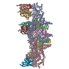
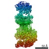
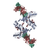
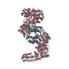
- PDB-6thh: Crystal structure of type I-D CRISPR-Cas nuclease Cas10d in compl... -
+
Open data
ID or keywords:
Loading...
-
Basic information
Entry
Database: PDB / ID: 6thh
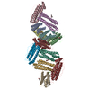
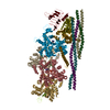
Title
Crystal structure of type I-D CRISPR-Cas nuclease Cas10d in complex with the SIRV3 AcrID1 (gp02) anti-CRISPR protein
Components
CRISPR-associated protein, CscA
SIRV3 AcrID1 (gp02) anti-CRISPR protein
Keywords
IMMUNE SYSTEM / CRISPR / Cas / type I-D / Cas10 / Cas10d / complex / anti-CRISPR
Function / homology
Sulfolobus islandicus filamentous virus, Orf14 / Protein of unknown function (DUF1374) / metal ion binding / PHOSPHATE ION / Uncharacterized protein / CRISPR-associated protein, CscA
Protocol: SINGLE WAVELENGTH / Monochromatic (M) / Laue (L): M / Scattering type: x-ray
Radiation wavelength
Wavelength: 0.9793 Å / Relative weight: 1
Reflection
Resolution: 3.48→67.542 Å / Num. obs: 21713 / % possible obs: 99.9 % / Redundancy: 2 % / Biso Wilson estimate: 137.1 Å2 / CC1/2: 0.99 / Rmerge(I) obs: 0.06 / Net I/σ(I): 11.9
Reflection shell
Resolution: 3.48→3.6 Å / Redundancy: 2 % / Rmerge(I) obs: 0.68 / Mean I/σ(I) obs: 1.2 / Num. unique obs: 2139 / CC1/2: 0.6 / % possible all: 99.8
-
Phasing
Phasing
Method: SAD
-
Processing
Software
Name
Version
Classification
XDS
datareduction
SHELXDE
phasing
PHENIX
1.16_3549
refinement
PDB_EXTRACT
3.25
dataextraction
XSCALE
datascaling
Refinement
Method to determine structure: SAD / Resolution: 3.48→67.542 Å / SU ML: 0.46 / Cross valid method: THROUGHOUT / σ(F): 1.34 / Phase error: 26.77 / Stereochemistry target values: ML
Rfactor
Num. reflection
% reflection
Rfree
0.2538
1841
8.49 %
Rwork
0.2364
19855
-
obs
0.2379
21696
99.88 %
Solvent computation
Shrinkage radii: 0.9 Å / VDW probe radii: 1.11 Å / Solvent model: FLAT BULK SOLVENT MODEL
In the structure databanks used in Yorodumi, some data are registered as the other names, "COVID-19 virus" and "2019-nCoV". Here are the details of the virus and the list of structure data.
Jan 31, 2019. EMDB accession codes are about to change! (news from PDBe EMDB page)
EMDB accession codes are about to change! (news from PDBe EMDB page)
The allocation of 4 digits for EMDB accession codes will soon come to an end. Whilst these codes will remain in use, new EMDB accession codes will include an additional digit and will expand incrementally as the available range of codes is exhausted. The current 4-digit format prefixed with “EMD-” (i.e. EMD-XXXX) will advance to a 5-digit format (i.e. EMD-XXXXX), and so on. It is currently estimated that the 4-digit codes will be depleted around Spring 2019, at which point the 5-digit format will come into force.
The EM Navigator/Yorodumi systems omit the EMD- prefix.
Related info.:Q: What is EMD? / ID/Accession-code notation in Yorodumi/EM Navigator
Yorodumi is a browser for structure data from EMDB, PDB, SASBDB, etc.
This page is also the successor to EM Navigator detail page, and also detail information page/front-end page for Omokage search.
The word "yorodu" (or yorozu) is an old Japanese word meaning "ten thousand". "mi" (miru) is to see.
Related info.:EMDB / PDB / SASBDB / Comparison of 3 databanks / Yorodumi Search / Aug 31, 2016. New EM Navigator & Yorodumi / Yorodumi Papers / Jmol/JSmol / Function and homology information / Changes in new EM Navigator and Yorodumi
 Movie
Movie Controller

 Yorodumi
 Open data
 Basic information
 Components
 Keywords
 Function and homology information
 Sulfolobus islandicus rudivirus 3
 X-RAY DIFFRACTION /
  Authors
 Denmark, 2items
  Citation
 Structure visualization
 Downloads & links
 Other downloads
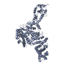


 PDBj
PDBj
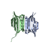
 Assembly
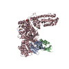


 Mass: 94.971 Da / Num. of mol.: 1 / Source method: obtained synthetically / Formula: PO4 / Feature type: SUBJECT OF INVESTIGATION
 Mass: 65.409 Da / Num. of mol.: 1 / Source method: obtained synthetically / Formula: Zn / Feature type: SUBJECT OF INVESTIGATION
 Sample preparation
 / Beamline: P14 (MX2) / Wavelength: 0.9793 Å
 Processing