 Movie
Movie Controller

[English] 日本語
 Yorodumi
- PDB-6tgi: Crystal structure of VIM-2 in complex with triazole-based inhibit... -
+ Open data

- Basic information
| Entry | Database: PDB / ID: 6tgi | ||||||
|---|---|---|---|---|---|---|---|
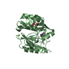
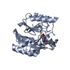
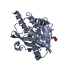
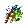
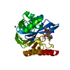
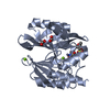
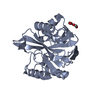
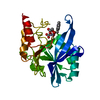
| Title | Crystal structure of VIM-2 in complex with triazole-based inhibitor OP24 | ||||||
 Components Components | Vim-1 | ||||||
 Keywords Keywords | HYDROLASE / New Delhi metallo-beta-lactamase | ||||||
| Function / homology |  Function and homology information Function and homology informationmembrane => GO:0016020 / antibiotic catabolic process / beta-lactamase activity / beta-lactamase / periplasmic space / response to antibiotic / metal ion binding Similarity search - Function | ||||||
| Biological species |   Pseudomonas aeruginosa (bacteria) Pseudomonas aeruginosa (bacteria) | ||||||
| Method |  X-RAY DIFFRACTION / SYNCHROTRON / MOLECULAR REPLACEMENT / Resolution: 1.6 Å X-RAY DIFFRACTION / SYNCHROTRON / MOLECULAR REPLACEMENT / Resolution: 1.6 Å | ||||||
 Authors Authors | Maso, L. / Spirakis, F. / Santucci, M. / Simon, C. / Docquier, J.D. / Cruciani, G. / Costi, M.P. / Tondi, D. / Cendron, L. | ||||||
| Funding support |  Italy, 1items Italy, 1items
| ||||||
 Citation Citation | Journal: Sci Rep / Year: 2020 Title: Virtual screening identifies broad-spectrum beta-lactamase inhibitors with activity on clinically relevant serine- and metallo-carbapenemases. Authors: Spyrakis, F. / Santucci, M. / Maso, L. / Cross, S. / Gianquinto, E. / Sannio, F. / Verdirosa, F. / De Luca, F. / Docquier, J.D. / Cendron, L. / Tondi, D. / Venturelli, A. / Cruciani, G. / Costi, M.P. | ||||||
| History |
|
- Structure visualization
| Structure viewer | Molecule: MolmilJmol/JSmol |
|---|
- Downloads & links
-Download
| PDBx/mmCIF format | 6tgi.cif.gz | 114.6 KB | Display | PDBx/mmCIF format |
|---|---|---|---|---|
| PDB format | pdb6tgi.ent.gz | 87.1 KB | Display | PDB format |
| PDBx/mmJSON format | 6tgi.json.gz | Tree view | PDBx/mmJSON format | |
| Others |  Other downloads Other downloads |
-Validation report
| Arichive directory | https://data.pdbj.org/pub/pdb/validation_reports/tg/6tgiftp://data.pdbj.org/pub/pdb/validation_reports/tg/6tgi | HTTPS FTP |
|---|
-Related structure data
| Related structure data | 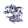 6tgdC  4bz3S S: Starting model for refinement C: citing same article ( |
|---|---|
| Similar structure data |


-Links
 PDBj
PDBj

- Assembly
| Deposited unit | 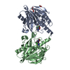
| ||||||||
|---|---|---|---|---|---|---|---|---|---|
| 1 | 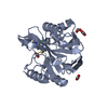
| ||||||||
| 2 | 
| ||||||||
| Unit cell |
|
-Components
| #1: Protein | Mass: 26142.004 Da / Num. of mol.: 2 Source method: isolated from a genetically manipulated source Details: Missing residues are present in the crystallized protein but not visible in the electron density maps. Source: (gene. exp.) Pseudomonas aeruginosa (bacteria) / Gene: blm, NCTC13437_04762 / Production host: References: UniProt: A0A485HTJ5, UniProt: Q9K2N0*PLUS, beta-lactamase #2: Chemical | ChemComp-ZN /   Mass: 65.409 Da / Num. of mol.: 6 / Source method: obtained synthetically / Formula: Zn Mass: 65.409 Da / Num. of mol.: 6 / Source method: obtained synthetically / Formula: Zn#3: Chemical | ChemComp-FMT /   Mass: 46.025 Da / Num. of mol.: 8 / Source method: obtained synthetically / Formula: CH2O2 Mass: 46.025 Da / Num. of mol.: 8 / Source method: obtained synthetically / Formula: CH2O2#4: Chemical |   Mass: 257.743 Da / Num. of mol.: 2 / Source method: obtained synthetically / Formula: C9H12ClN5S / Feature type: SUBJECT OF INVESTIGATION Mass: 257.743 Da / Num. of mol.: 2 / Source method: obtained synthetically / Formula: C9H12ClN5S / Feature type: SUBJECT OF INVESTIGATION#5: Water | ChemComp-HOH / |  Mass: 18.015 Da / Num. of mol.: 507 / Source method: isolated from a natural source / Formula: H2O Mass: 18.015 Da / Num. of mol.: 507 / Source method: isolated from a natural source / Formula: H2OHas ligand of interest | Y | |
|---|
-Experimental details
-Experiment
| Experiment | Method: X-RAY DIFFRACTION / Number of used crystals: 1 |
|---|
- Sample preparation
| Crystal | Density Matthews: 2.01 Å3/Da / Density % sol: 38.84 % |
|---|---|
| Crystal grow | Temperature: 293 K / Method: vapor diffusion, sitting drop / Details: 0.25 M Magnesium Formate, 27 % PEG3350 |
-Data collection
| Diffraction | Mean temperature: 100 K / Serial crystal experiment: N |
|---|---|
| Diffraction source | Source: SYNCHROTRON / Site: SLS  / Beamline: X06DA / Wavelength: 1.001 Å / Beamline: X06DA / Wavelength: 1.001 Å |
| Detector | Type: DECTRIS PILATUS 2M-F / Detector: PIXEL / Date: Mar 25, 2019 |
| Radiation | Protocol: SINGLE WAVELENGTH / Monochromatic (M) / Laue (L): M / Scattering type: x-ray |
| Radiation wavelength | Wavelength: 1.001 Å / Relative weight: 1 |
| Reflection | Resolution: 1.6→39.56 Å / Num. obs: 54492 / % possible obs: 97.4 % / Redundancy: 3.8 % / Biso Wilson estimate: 17.3 Å2 / Rmerge(I) obs: 0.084 / Net I/σ(I): 8.2 |
| Reflection shell | Resolution: 1.6→1.66 Å / Rmerge(I) obs: 0.553 / Mean I/σ(I) obs: 1.9 / Num. unique obs: 5436 |
- Processing
| Software |
| |||||||||||||||||||||||||||||||||||||||||||||||||||||||||||||||||||||||||||||||||||||||||||||||||||||||||||||||||||||||||||||||||||||||||||||||||
|---|---|---|---|---|---|---|---|---|---|---|---|---|---|---|---|---|---|---|---|---|---|---|---|---|---|---|---|---|---|---|---|---|---|---|---|---|---|---|---|---|---|---|---|---|---|---|---|---|---|---|---|---|---|---|---|---|---|---|---|---|---|---|---|---|---|---|---|---|---|---|---|---|---|---|---|---|---|---|---|---|---|---|---|---|---|---|---|---|---|---|---|---|---|---|---|---|---|---|---|---|---|---|---|---|---|---|---|---|---|---|---|---|---|---|---|---|---|---|---|---|---|---|---|---|---|---|---|---|---|---|---|---|---|---|---|---|---|---|---|---|---|---|---|---|---|---|
| Refinement | Method to determine structure: MOLECULAR REPLACEMENT Starting model: 4BZ3 Resolution: 1.6→39.56 Å / Cor.coef. Fo:Fc: 0.965 / Cor.coef. Fo:Fc free: 0.947 / Cross valid method: THROUGHOUT / σ(F): 0 / ESU R: 0.099 / ESU R Free: 0.101 / Details: U VALUES : REFINED INDIVIDUALLY
| |||||||||||||||||||||||||||||||||||||||||||||||||||||||||||||||||||||||||||||||||||||||||||||||||||||||||||||||||||||||||||||||||||||||||||||||||
| Solvent computation | Ion probe radii: 0.8 Å / Shrinkage radii: 0.8 Å / VDW probe radii: 1.2 Å | |||||||||||||||||||||||||||||||||||||||||||||||||||||||||||||||||||||||||||||||||||||||||||||||||||||||||||||||||||||||||||||||||||||||||||||||||
| Displacement parameters | Biso max: 72.09 Å2 / Biso mean: 21.35 Å2 / Biso min: 9.36 Å2
| |||||||||||||||||||||||||||||||||||||||||||||||||||||||||||||||||||||||||||||||||||||||||||||||||||||||||||||||||||||||||||||||||||||||||||||||||
| Refinement step | Cycle: final / Resolution: 1.6→39.56 Å
| |||||||||||||||||||||||||||||||||||||||||||||||||||||||||||||||||||||||||||||||||||||||||||||||||||||||||||||||||||||||||||||||||||||||||||||||||
| Refine LS restraints |
| |||||||||||||||||||||||||||||||||||||||||||||||||||||||||||||||||||||||||||||||||||||||||||||||||||||||||||||||||||||||||||||||||||||||||||||||||
| LS refinement shell | Resolution: 1.6→1.64 Å / Rfactor Rfree error: 0 / Total num. of bins used: 20
|
