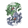
 Movie
Movie Controller

[English] 日本語
 Yorodumi
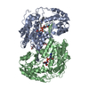
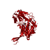
- PDB-6te5: Crystal structure of human Aldehyde dehydrogenase 1A3 in complex ... -
+ Open data

- Basic information
| Entry | Database: PDB / ID: 6te5 | ||||||
|---|---|---|---|---|---|---|---|
| Title | Crystal structure of human Aldehyde dehydrogenase 1A3 in complex with LQ43 inhibitor compound | ||||||
 Components Components | Aldehyde dehydrogenase family 1 member A3 | ||||||
 Keywords Keywords | OXIDOREDUCTASE / Dehydrogenase isoenzyme ALDH1A3 Selective Inhibitor High grade Glioma | ||||||
| Function / homology |  Function and homology information Function and homology informationnucleus accumbens development / optic cup morphogenesis involved in camera-type eye development / olfactory pit development / Harderian gland development / embryonic eye morphogenesis / retinoic acid biosynthetic process / retinal dehydrogenase / embryonic camera-type eye development / thyroid hormone binding / aldehyde dehydrogenase [NAD(P)+] activity ...nucleus accumbens development / optic cup morphogenesis involved in camera-type eye development / olfactory pit development / Harderian gland development / embryonic eye morphogenesis / retinoic acid biosynthetic process / retinal dehydrogenase / embryonic camera-type eye development / thyroid hormone binding / aldehyde dehydrogenase [NAD(P)+] activity / RA biosynthesis pathway / aldehyde metabolic process / righting reflex / retinal metabolic process / Developmental Lineage of Mammary Stem Cells / aldehyde dehydrogenase (NAD+) activity / retinal dehydrogenase (NAD+) activity / retinoic acid metabolic process / retinol metabolic process / neuromuscular process controlling balance / inner ear morphogenesis / face development / Developmental Lineage of Mammary Gland Myoepithelial Cells / NAD+ binding / locomotory behavior / protein homotetramerization / positive regulation of apoptotic process / apoptotic process / protein homodimerization activity / extracellular exosome / cytoplasm / cytosol Similarity search - Function | ||||||
| Biological species |  Homo sapiens (human) Homo sapiens (human) | ||||||
| Method |  X-RAY DIFFRACTION / SYNCHROTRON / MOLECULAR REPLACEMENT / Resolution: 3.25 Å X-RAY DIFFRACTION / SYNCHROTRON / MOLECULAR REPLACEMENT / Resolution: 3.25 Å | ||||||
 Authors Authors | Gelardi, E.L.M. / Garavaglia, S. | ||||||
| Funding support |  Italy, 1items Italy, 1items
| ||||||
 Citation Citation | Journal: J.Med.Chem. / Year: 2020 Title: Imidazo[1,2- a ]pyridine Derivatives as Aldehyde Dehydrogenase Inhibitors: Novel Chemotypes to Target Glioblastoma Stem Cells. Authors: Quattrini, L. / Gelardi, E.L.M. / Coviello, V. / Sartini, S. / Ferraris, D.M. / Mori, M. / Nakano, I. / Garavaglia, S. / La Motta, C. | ||||||
| History |
|
- Structure visualization
| Structure viewer | Molecule: MolmilJmol/JSmol |
|---|
- Downloads & links
-Download
| PDBx/mmCIF format | 6te5.cif.gz | 199.7 KB | Display | PDBx/mmCIF format |
|---|---|---|---|---|
| PDB format | pdb6te5.ent.gz | 158.5 KB | Display | PDB format |
| PDBx/mmJSON format | 6te5.json.gz | Tree view | PDBx/mmJSON format | |
| Others |  Other downloads Other downloads |
-Validation report
| Arichive directory | https://data.pdbj.org/pub/pdb/validation_reports/te/6te5ftp://data.pdbj.org/pub/pdb/validation_reports/te/6te5 | HTTPS FTP |
|---|
-Related structure data
| Related structure data | 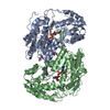 6s6wC 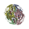 5fhzS C: citing same article ( S: Starting model for refinement |
|---|---|
| Similar structure data |


-Links
 PDBj
PDBj


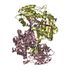
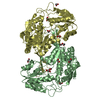
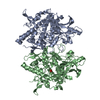
- Assembly
| Deposited unit | 
| ||||||||
|---|---|---|---|---|---|---|---|---|---|
| 1 |
| ||||||||
| Unit cell |
|
-Components
| #1: Protein | Mass: 56177.289 Da / Num. of mol.: 2 Source method: isolated from a genetically manipulated source Source: (gene. exp.) Homo sapiens (human) / Gene: ALDH1A3, ALDH6Production host:  References: UniProt: P47895, retinal dehydrogenase #2: Chemical |   Mass: 663.425 Da / Num. of mol.: 2 / Source method: obtained synthetically / Formula: C21H27N7O14P2 / Comment: NAD*YM Mass: 663.425 Da / Num. of mol.: 2 / Source method: obtained synthetically / Formula: C21H27N7O14P2 / Comment: NAD*YM#3: Chemical |   Mass: 360.406 Da / Num. of mol.: 2 / Source method: obtained synthetically / Formula: C22H20N2O3 / Feature type: SUBJECT OF INVESTIGATION Mass: 360.406 Da / Num. of mol.: 2 / Source method: obtained synthetically / Formula: C22H20N2O3 / Feature type: SUBJECT OF INVESTIGATION#4: Chemical | ChemComp-GOL / |   Mass: 92.094 Da / Num. of mol.: 1 / Source method: obtained synthetically / Formula: C3H8O3 Mass: 92.094 Da / Num. of mol.: 1 / Source method: obtained synthetically / Formula: C3H8O3#5: Water | ChemComp-HOH / |  Mass: 18.015 Da / Num. of mol.: 20 / Source method: isolated from a natural source / Formula: H2O Mass: 18.015 Da / Num. of mol.: 20 / Source method: isolated from a natural source / Formula: H2OHas ligand of interest | Y | Has protein modification | N | |
|---|
-Experimental details
-Experiment
| Experiment | Method: X-RAY DIFFRACTION / Number of used crystals: 1 |
|---|
- Sample preparation
| Crystal | Density Matthews: 2.57 Å3/Da / Density % sol: 52.13 % |
|---|---|
| Crystal grow | Temperature: 293.15 K / Method: vapor diffusion / Details: Sodium Malonate 2.4M pH 7.0 |
-Data collection
| Diffraction | Mean temperature: 100 K / Serial crystal experiment: N | |||||||||||||||||||||||||||||||||||||||||||||||||||||||||||||||||||||||||||||||||||||||||||||||||||||||||||||||||||||||||
|---|---|---|---|---|---|---|---|---|---|---|---|---|---|---|---|---|---|---|---|---|---|---|---|---|---|---|---|---|---|---|---|---|---|---|---|---|---|---|---|---|---|---|---|---|---|---|---|---|---|---|---|---|---|---|---|---|---|---|---|---|---|---|---|---|---|---|---|---|---|---|---|---|---|---|---|---|---|---|---|---|---|---|---|---|---|---|---|---|---|---|---|---|---|---|---|---|---|---|---|---|---|---|---|---|---|---|---|---|---|---|---|---|---|---|---|---|---|---|---|---|---|---|
| Diffraction source | Source: SYNCHROTRON / Site: ESRF  / Beamline: ID29 / Wavelength: 0.967 Å / Beamline: ID29 / Wavelength: 0.967 Å | |||||||||||||||||||||||||||||||||||||||||||||||||||||||||||||||||||||||||||||||||||||||||||||||||||||||||||||||||||||||||
| Detector | Type: DECTRIS PILATUS 6M / Detector: PIXEL / Date: Sep 30, 2018 | |||||||||||||||||||||||||||||||||||||||||||||||||||||||||||||||||||||||||||||||||||||||||||||||||||||||||||||||||||||||||
| Radiation | Protocol: SINGLE WAVELENGTH / Monochromatic (M) / Laue (L): M / Scattering type: x-ray | |||||||||||||||||||||||||||||||||||||||||||||||||||||||||||||||||||||||||||||||||||||||||||||||||||||||||||||||||||||||||
| Radiation wavelength | Wavelength: 0.967 Å / Relative weight: 1 | |||||||||||||||||||||||||||||||||||||||||||||||||||||||||||||||||||||||||||||||||||||||||||||||||||||||||||||||||||||||||
| Reflection | Resolution: 3.25→47.956 Å / Num. all: 18856 / Num. obs: 18856 / % possible obs: 99.9 % / Redundancy: 5.6 % / Rpim(I) all: 0.076 / Rrim(I) all: 0.185 / Rsym value: 0.168 / Net I/av σ(I): 4.3 / Net I/σ(I): 8.9 / Num. measured all: 105886 | |||||||||||||||||||||||||||||||||||||||||||||||||||||||||||||||||||||||||||||||||||||||||||||||||||||||||||||||||||||||||
| Reflection shell | Diffraction-ID: 1
|
- Processing
| Software |
| ||||||||||||||||||
|---|---|---|---|---|---|---|---|---|---|---|---|---|---|---|---|---|---|---|---|
| Refinement | Method to determine structure: MOLECULAR REPLACEMENT Starting model: 5FHZ Resolution: 3.25→47.956 Å / Cross valid method: THROUGHOUT
| ||||||||||||||||||
| Displacement parameters | Biso max: 138.55 Å2 / Biso mean: 57.2162 Å2 / Biso min: 22.26 Å2 | ||||||||||||||||||
| Refinement step | Cycle: LAST / Resolution: 3.25→47.956 Å
|
