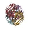
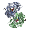
- PDB-4f3x: Crystal structure of putative aldehyde dehydrogenase from Sinorhi... -
+
Open data
ID or keywords:
Loading...
-
Basic information
Entry
Database: PDB / ID: 4f3x
Title
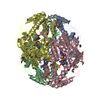
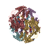
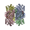
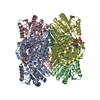
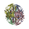
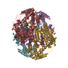
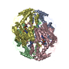
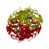
Crystal structure of putative aldehyde dehydrogenase from Sinorhizobium meliloti 1021 complexed with NAD
Components
Putative aldehyde dehydrogenase
Keywords
OXIDOREDUCTASE / STRUCTURAL GENOMICS / PROTEIN STRUCTURE INITIATIVE / NYSGRC / PSI-Biology / New York Structural Genomics Research Consortium
Function / homology
Function and homology information
aldehyde dehydrogenase [NAD(P)+] activity / Oxidoreductases; Acting on the aldehyde or oxo group of donors; With NAD+ or NADP+ as acceptor / nucleotide binding Similarity search - Function
Aminobutyraldehyde dehydrogenase / Aldehyde Dehydrogenase; Chain A, domain 2 / Aldehyde Dehydrogenase; Chain A, domain 2 / Aldehyde Dehydrogenase; Chain A, domain 1 / Aldehyde Dehydrogenase; Chain A, domain 1 / Aldehyde dehydrogenase, glutamic acid active site / Aldehyde dehydrogenases glutamic acid active site. / Aldehyde dehydrogenase domain / Aldehyde dehydrogenase family / Aldehyde dehydrogenase, C-terminal ...Aminobutyraldehyde dehydrogenase / Aldehyde Dehydrogenase; Chain A, domain 2 / Aldehyde Dehydrogenase; Chain A, domain 2 / Aldehyde Dehydrogenase; Chain A, domain 1 / Aldehyde Dehydrogenase; Chain A, domain 1 / Aldehyde dehydrogenase, glutamic acid active site / Aldehyde dehydrogenases glutamic acid active site. / Aldehyde dehydrogenase domain / Aldehyde dehydrogenase family / Aldehyde dehydrogenase, C-terminal / Aldehyde dehydrogenase, N-terminal / Aldehyde/histidinol dehydrogenase / 3-Layer(aba) Sandwich / Alpha Beta Similarity search - Domain/homology
Mass: 54532.391 Da / Num. of mol.: 4 Source method: isolated from a genetically manipulated source Source: (gene. exp.) Sinorhizobium meliloti (bacteria) / Strain: 1021 / Gene: R02268, SMc01656 / Plasmid: BC-PSGX3(BC) / Production host: Escherichia coli (E. coli) / Strain (production host): BL21(DE3)CODON+RIL References: UniProt: Q92ND9, Oxidoreductases; Acting on the aldehyde or oxo group of donors; With NAD+ or NADP+ as acceptor
Resolution: 2.01→19.91 Å / Cor.coef. Fo:Fc: 0.972 / Cor.coef. Fo:Fc free: 0.951 / WRfactor Rfree: 0.2121 / WRfactor Rwork: 0.1637 / Occupancy max: 1 / Occupancy min: 0.5 / FOM work R set: 0.8078 / SU B: 11.607 / SU ML: 0.147 / SU R Cruickshank DPI: 0.1903 / SU Rfree: 0.1721 / Cross valid method: THROUGHOUT / σ(F): 0 / ESU R: 0.19 / ESU R Free: 0.172 / Stereochemistry target values: MAXIMUM LIKELIHOOD Details: U VALUES : WITH TLS ADDED HYDROGENS HAVE BEEN USED IF PRESENT IN THE INPUT
Rfactor
Num. reflection
% reflection
Selection details
Rfree
0.2326
6378
5 %
RANDOM
Rwork
0.1802
-
-
-
obs
0.1828
126673
95.55 %
-
Solvent computation
Ion probe radii: 0.8 Å / Shrinkage radii: 0.8 Å / VDW probe radii: 1.2 Å / Solvent model: MASK
In the structure databanks used in Yorodumi, some data are registered as the other names, "COVID-19 virus" and "2019-nCoV". Here are the details of the virus and the list of structure data.
Jan 31, 2019. EMDB accession codes are about to change! (news from PDBe EMDB page)
EMDB accession codes are about to change! (news from PDBe EMDB page)
The allocation of 4 digits for EMDB accession codes will soon come to an end. Whilst these codes will remain in use, new EMDB accession codes will include an additional digit and will expand incrementally as the available range of codes is exhausted. The current 4-digit format prefixed with “EMD-” (i.e. EMD-XXXX) will advance to a 5-digit format (i.e. EMD-XXXXX), and so on. It is currently estimated that the 4-digit codes will be depleted around Spring 2019, at which point the 5-digit format will come into force.
The EM Navigator/Yorodumi systems omit the EMD- prefix.
Related info.:Q: What is EMD? / ID/Accession-code notation in Yorodumi/EM Navigator
Yorodumi is a browser for structure data from EMDB, PDB, SASBDB, etc.
This page is also the successor to EM Navigator detail page, and also detail information page/front-end page for Omokage search.
The word "yorodu" (or yorozu) is an old Japanese word meaning "ten thousand". "mi" (miru) is to see.
Related info.:EMDB / PDB / SASBDB / Comparison of 3 databanks / Yorodumi Search / Aug 31, 2016. New EM Navigator & Yorodumi / Yorodumi Papers / Jmol/JSmol / Function and homology information / Changes in new EM Navigator and Yorodumi
 Movie
Movie Controller

 Yorodumi
 Open data
 Basic information
 Components
 Keywords
 Function and homology information
 Sinorhizobium meliloti (bacteria)
 X-RAY DIFFRACTION /
  Authors
 Citation
 Structure visualization
 Downloads & links
 Other downloads

 PDBj
PDBj

 Assembly
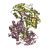

 Mass: 663.425 Da / Num. of mol.: 4 / Source method: obtained synthetically / Formula: C21H27N7O14P2 / Comment: NAD*YM
 Mass: 92.094 Da / Num. of mol.: 5 / Source method: obtained synthetically / Formula: C3H8O3
 Mass: 18.015 Da / Num. of mol.: 642 / Source method: isolated from a natural source / Formula: H2O
 Sample preparation
 / Beamline: X29A / Wavelength: 1.075 Å
 Processing