 Movie
Movie Controller

[English] 日本語
 Yorodumi
- PDB-6tdf: Crystal structure of Aspergillus fumigatus Glucosamine-6-phosphat... -
+ Open data

- Basic information
| Entry | Database: PDB / ID: 6tdf | ||||||
|---|---|---|---|---|---|---|---|
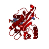
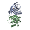
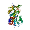
| Title | Crystal structure of Aspergillus fumigatus Glucosamine-6-phosphate N-acetyltransferase 1 in complex with compound 3 | ||||||
 Components Components | Glucosamine 6-phosphate N-acetyltransferase | ||||||
 Keywords Keywords | TRANSFERASE / fragment screening / anti fungal / Aspergillus fumigatus / inhibitor | ||||||
| Function / homology |  Function and homology information Function and homology informationglucosamine-phosphate N-acetyltransferase / glucosamine 6-phosphate N-acetyltransferase activity / UDP-N-acetylglucosamine biosynthetic process / endoplasmic reticulum membrane Similarity search - Function | ||||||
| Biological species |  | ||||||
| Method |  X-RAY DIFFRACTION / SYNCHROTRON / MOLECULAR REPLACEMENT / Resolution: 2.01 Å X-RAY DIFFRACTION / SYNCHROTRON / MOLECULAR REPLACEMENT / Resolution: 2.01 Å | ||||||
 Authors Authors | Raimi, O.G. / Stanley, M. / Lockhart, D. | ||||||
| Funding support |  United Kingdom, 1items United Kingdom, 1items
| ||||||
 Citation Citation | Journal: J.Biol.Chem. / Year: 2020 Title: Targeting a critical step in fungal hexosamine biosynthesis. Authors: Lockhart, D.E.A. / Stanley, M. / Raimi, O.G. / Robinson, D.A. / Boldovjakova, D. / Squair, D.R. / Ferenbach, A.T. / Fang, W. / van Aalten, D.M.F. | ||||||
| History |
|
- Structure visualization
| Structure viewer | Molecule: MolmilJmol/JSmol |
|---|
- Downloads & links
-Download
| PDBx/mmCIF format | 6tdf.cif.gz | 52.3 KB | Display | PDBx/mmCIF format |
|---|---|---|---|---|
| PDB format | pdb6tdf.ent.gz | 35.1 KB | Display | PDB format |
| PDBx/mmJSON format | 6tdf.json.gz | Tree view | PDBx/mmJSON format | |
| Others |  Other downloads Other downloads |
-Validation report
| Arichive directory | https://data.pdbj.org/pub/pdb/validation_reports/td/6tdfftp://data.pdbj.org/pub/pdb/validation_reports/td/6tdf | HTTPS FTP |
|---|
-Related structure data
| Related structure data |  6tdgC  6tdhC  2vezS S: Starting model for refinement C: citing same article ( |
|---|---|
| Similar structure data |


-Links
 PDBj
PDBj

- Assembly
| Deposited unit | 
| |||||||||||||||
|---|---|---|---|---|---|---|---|---|---|---|---|---|---|---|---|---|
| 1 | 
| |||||||||||||||
| Unit cell |
| |||||||||||||||
| Components on special symmetry positions |
|
-Components
| #1: Protein | Mass: 21126.113 Da / Num. of mol.: 1 Source method: isolated from a genetically manipulated source Source: (gene. exp.)  References: UniProt: Q4WCU5, glucosamine-phosphate N-acetyltransferase |
|---|---|
| #2: Chemical | ChemComp-ACO /   Mass: 809.571 Da / Num. of mol.: 1 / Source method: obtained synthetically / Formula: C23H38N7O17P3S Mass: 809.571 Da / Num. of mol.: 1 / Source method: obtained synthetically / Formula: C23H38N7O17P3S |
| #3: Chemical | ChemComp-N3Q /   Mass: 317.171 Da / Num. of mol.: 1 / Source method: obtained synthetically / Formula: C12H14Cl2N4O2 / Feature type: SUBJECT OF INVESTIGATION Mass: 317.171 Da / Num. of mol.: 1 / Source method: obtained synthetically / Formula: C12H14Cl2N4O2 / Feature type: SUBJECT OF INVESTIGATION |
| #4: Sugar | ChemComp-G6P /   Type: D-saccharide, alpha linking / Mass: 260.136 Da / Num. of mol.: 1 Type: D-saccharide, alpha linking / Mass: 260.136 Da / Num. of mol.: 1Source method: isolated from a genetically manipulated source Formula: C6H13O9P |
| #5: Water | ChemComp-HOH /  Mass: 18.015 Da / Num. of mol.: 45 / Source method: isolated from a natural source / Formula: H2O Mass: 18.015 Da / Num. of mol.: 45 / Source method: isolated from a natural source / Formula: H2O |
| Has ligand of interest | Y |
-Experimental details
-Experiment
| Experiment | Method: X-RAY DIFFRACTION / Number of used crystals: 1 |
|---|
- Sample preparation
| Crystal | Density Matthews: 2.7 Å3/Da / Density % sol: 54.41 % |
|---|---|
| Crystal grow | Temperature: 277 K / Method: vapor diffusion, sitting drop / Details: 1. 10% peg 1000, 10%peg 8000 2. 30% peg1500 |
-Data collection
| Diffraction | Mean temperature: 277 K / Serial crystal experiment: N |
|---|---|
| Diffraction source | Source: SYNCHROTRON / Site: ESRF  / Beamline: ID30B / Wavelength: 0.966 Å / Beamline: ID30B / Wavelength: 0.966 Å |
| Detector | Type: DECTRIS PILATUS3 2M / Detector: PIXEL / Date: Nov 17, 2017 |
| Radiation | Protocol: SINGLE WAVELENGTH / Monochromatic (M) / Laue (L): M / Scattering type: x-ray |
| Radiation wavelength | Wavelength: 0.966 Å / Relative weight: 1 |
| Reflection | Resolution: 2→58.2 Å / Num. obs: 13064 / % possible obs: 94.6 % / Redundancy: 4 % / CC1/2: 1 / Rsym value: 0.049 / Net I/σ(I): 14.5 |
| Reflection shell | Resolution: 2→2.08 Å / Redundancy: 4.2 % / Mean I/σ(I) obs: 2.4 / Num. unique obs: 13064 / CC1/2: 1 / Rsym value: 0.578 |
- Processing
| Software |
| ||||||||||||||||||||||||||||||||||||||||||||||||||||||||||||
|---|---|---|---|---|---|---|---|---|---|---|---|---|---|---|---|---|---|---|---|---|---|---|---|---|---|---|---|---|---|---|---|---|---|---|---|---|---|---|---|---|---|---|---|---|---|---|---|---|---|---|---|---|---|---|---|---|---|---|---|---|---|
| Refinement | Method to determine structure: MOLECULAR REPLACEMENT Starting model: 2vez Resolution: 2.01→58.2 Å / Cor.coef. Fo:Fc: 0.961 / Cor.coef. Fo:Fc free: 0.945 / SU B: 7.049 / SU ML: 0.179 / Cross valid method: THROUGHOUT / σ(F): 0 / ESU R: 0.203 / ESU R Free: 0.187 / Stereochemistry target values: MAXIMUM LIKELIHOOD Details: HYDROGENS HAVE BEEN ADDED IN THE RIDING POSITIONS U VALUES : REFINED INDIVIDUALLY
| ||||||||||||||||||||||||||||||||||||||||||||||||||||||||||||
| Solvent computation | Ion probe radii: 0.8 Å / Shrinkage radii: 0.8 Å / VDW probe radii: 1.2 Å / Solvent model: MASK | ||||||||||||||||||||||||||||||||||||||||||||||||||||||||||||
| Displacement parameters | Biso max: 90.66 Å2 / Biso mean: 43.114 Å2 / Biso min: 29.03 Å2
| ||||||||||||||||||||||||||||||||||||||||||||||||||||||||||||
| Refinement step | Cycle: final / Resolution: 2.01→58.2 Å
| ||||||||||||||||||||||||||||||||||||||||||||||||||||||||||||
| Refine LS restraints |
| ||||||||||||||||||||||||||||||||||||||||||||||||||||||||||||
| LS refinement shell | Resolution: 2.01→2.08 Å / Rfactor Rfree error: 0
|
