 Movie
Movie Controller

[English] 日本語
 Yorodumi
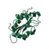
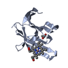
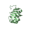
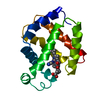
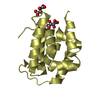
- PDB-6td7: Structure of truncated hemoglobin THB11 from Chlamydomonas reinhardtii -
+ Open data

- Basic information
| Entry | Database: PDB / ID: 6td7 | |||||||||
|---|---|---|---|---|---|---|---|---|---|---|
| Title | Structure of truncated hemoglobin THB11 from Chlamydomonas reinhardtii | |||||||||
 Components Components | THB11 | |||||||||
 Keywords Keywords | OXYGEN BINDING / heme binding / truncated hemoglobin / nitrite reduction / pentacoordination | |||||||||
| Function / homology |  Function and homology information Function and homology informationthioredoxin peroxidase activity / cell redox homeostasis / oxygen binding / cellular response to oxidative stress / heme binding / metal ion binding / cytoplasm Similarity search - Function | |||||||||
| Biological species |   Chlamydomonas reinhardtii (plant) Chlamydomonas reinhardtii (plant) | |||||||||
| Method |  X-RAY DIFFRACTION / SYNCHROTRON / SAD / Resolution: 1.75 Å X-RAY DIFFRACTION / SYNCHROTRON / SAD / Resolution: 1.75 Å | |||||||||
 Authors Authors | Huwald, D. / Gasper, R. / Hemschemeier, A. / Hofmann, E. | |||||||||
| Funding support |  Germany, 2items Germany, 2items
| |||||||||
 Citation Citation | Journal: J.Biol.Inorg.Chem. / Year: 2020 Title: Distinctive structural properties of THB11, a pentacoordinate Chlamydomonas reinhardtii truncated hemoglobin with N- and C-terminal extensions. Authors: Huwald, D. / Duda, S. / Gasper, R. / Olieric, V. / Hofmann, E. / Hemschemeier, A. | |||||||||
| History |
|
- Structure visualization
| Structure viewer | Molecule: MolmilJmol/JSmol |
|---|
- Downloads & links
-Download
| PDBx/mmCIF format | 6td7.cif.gz | 82.8 KB | Display | PDBx/mmCIF format |
|---|---|---|---|---|
| PDB format | pdb6td7.ent.gz | 51.2 KB | Display | PDB format |
| PDBx/mmJSON format | 6td7.json.gz | Tree view | PDBx/mmJSON format | |
| Others |  Other downloads Other downloads |
-Validation report
| Arichive directory | https://data.pdbj.org/pub/pdb/validation_reports/td/6td7ftp://data.pdbj.org/pub/pdb/validation_reports/td/6td7 | HTTPS FTP |
|---|
-Related structure data
| Similar structure data |
|---|


-Links
 PDBj
PDBj


- Assembly
| Deposited unit | 
| ||||||||||||
|---|---|---|---|---|---|---|---|---|---|---|---|---|---|
| 1 |
| ||||||||||||
| Unit cell |
|
-Components
| #1: Protein | Mass: 15865.302 Da / Num. of mol.: 1 Source method: isolated from a genetically manipulated source Source: (gene. exp.) Chlamydomonas reinhardtii (plant) / Gene: CHLRE_16g662750v5 / Production host:  |
|---|---|
| #2: Chemical | ChemComp-HEM /   Mass: 616.487 Da / Num. of mol.: 1 / Source method: obtained synthetically / Formula: C34H32FeN4O4 / Feature type: SUBJECT OF INVESTIGATION Mass: 616.487 Da / Num. of mol.: 1 / Source method: obtained synthetically / Formula: C34H32FeN4O4 / Feature type: SUBJECT OF INVESTIGATION |
| #3: Chemical | ChemComp-CYN /   Mass: 26.017 Da / Num. of mol.: 1 / Source method: obtained synthetically / Formula: CN / Feature type: SUBJECT OF INVESTIGATION Mass: 26.017 Da / Num. of mol.: 1 / Source method: obtained synthetically / Formula: CN / Feature type: SUBJECT OF INVESTIGATION |
| #4: Water | ChemComp-HOH /  Mass: 18.015 Da / Num. of mol.: 153 / Source method: isolated from a natural source / Formula: H2O Mass: 18.015 Da / Num. of mol.: 153 / Source method: isolated from a natural source / Formula: H2O |
| Has ligand of interest | Y |
-Experimental details
-Experiment
| Experiment | Method: X-RAY DIFFRACTION / Number of used crystals: 1 |
|---|
- Sample preparation
| Crystal | Density Matthews: 2.07 Å3/Da / Density % sol: 40.6 % |
|---|---|
| Crystal grow | Temperature: 277 K / Method: vapor diffusion, sitting drop / pH: 7 Details: protein 19 mg*ml-1 in 15 mM Tris-HCl pH 8.0 mixed 1:1 with 0.1 M HEPES pH 7.0, 21% PEG 6.000 |
-Data collection
| Diffraction | Mean temperature: 100 K / Serial crystal experiment: N |
|---|---|
| Diffraction source | Source: SYNCHROTRON / Site: SLS  / Beamline: X10SA / Wavelength: 1.739 Å / Beamline: X10SA / Wavelength: 1.739 Å |
| Detector | Type: DECTRIS PILATUS 2M / Detector: PIXEL / Date: Mar 19, 2017 |
| Radiation | Protocol: SINGLE WAVELENGTH / Monochromatic (M) / Laue (L): M / Scattering type: x-ray |
| Radiation wavelength | Wavelength: 1.739 Å / Relative weight: 1 |
| Reflection | Resolution: 1.75→38.54 Å / Num. all: 594012 / Num. obs: 11933 / % possible obs: 94.26 % / Redundancy: 49.8 % / Biso Wilson estimate: 17.99 Å2 / CC1/2: 1 / Rmerge(I) obs: 0.09463 / Rpim(I) all: 0.01261 / Rrim(I) all: 0.09552 / Net I/σ(I): 38.39 |
| Reflection shell | Resolution: 1.75→1.813 Å / Redundancy: 6.2 % / Rmerge(I) obs: 0.5662 / Mean I/σ(I) obs: 2.64 / Num. unique all: 4429 / Num. unique obs: 711 / CC1/2: 0.83 / Rpim(I) all: 0.2366 / Rrim(I) all: 0.6174 / % possible all: 58.37 |
- Processing
| Software |
| ||||||||||||||||||||||||||||||||||||||||||||||||||||||||||||||||||||||
|---|---|---|---|---|---|---|---|---|---|---|---|---|---|---|---|---|---|---|---|---|---|---|---|---|---|---|---|---|---|---|---|---|---|---|---|---|---|---|---|---|---|---|---|---|---|---|---|---|---|---|---|---|---|---|---|---|---|---|---|---|---|---|---|---|---|---|---|---|---|---|---|
| Refinement | Method to determine structure: SAD / Resolution: 1.75→38.54 Å / SU ML: 0.1825 / Cross valid method: FREE R-VALUE / σ(F): 1.1 / Phase error: 19.6183
| ||||||||||||||||||||||||||||||||||||||||||||||||||||||||||||||||||||||
| Solvent computation | Shrinkage radii: 0.9 Å / VDW probe radii: 1.11 Å | ||||||||||||||||||||||||||||||||||||||||||||||||||||||||||||||||||||||
| Displacement parameters | Biso mean: 18.79 Å2 | ||||||||||||||||||||||||||||||||||||||||||||||||||||||||||||||||||||||
| Refinement step | Cycle: LAST / Resolution: 1.75→38.54 Å
| ||||||||||||||||||||||||||||||||||||||||||||||||||||||||||||||||||||||
| Refine LS restraints |
| ||||||||||||||||||||||||||||||||||||||||||||||||||||||||||||||||||||||
| LS refinement shell |
|
