[English] 日本語
Yorodumi
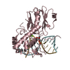
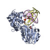
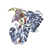
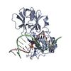
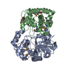
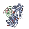
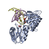
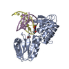
- PDB-6tc9: Crystal structure of MutM from Neisseria meningitidis -

+
Open data


ID or keywords:

Loading...

-
Basic information

Entry
Database: PDB / ID: 6tc9
TitleCrystal structure of MutM from Neisseria meningitidis
Components
  • DNA
  • DNA containing abasic site analogue
  • Formamidopyrimidine-DNA glycosylase
KeywordsDNA BINDING PROTEIN / MutM / Fpg/Nei / Neisseria meningitidis / BER / DNA repair
Function / homology
Function and homology information


oxidized purine nucleobase lesion DNA N-glycosylase activity / DNA-formamidopyrimidine glycosylase / class I DNA-(apurinic or apyrimidinic site) endonuclease activity / DNA-(apurinic or apyrimidinic site) lyase / base-excision repair / damaged DNA binding / zinc ion binding
Similarity search - Function
Formamidopyrimidine-DNA glycosylase / Zinc finger, DNA glycosylase/AP lyase-type / Zinc finger, FPG/IleRS-type / DNA glycosylase/AP lyase, zinc finger domain, DNA-binding site / Zinc finger found in FPG and IleRS / Zinc finger FPG-type signature. / Zinc finger FPG-type profile. / Formamidopyrimidine-DNA glycosylase H2TH domain / Formamidopyrimidine-DNA glycosylase N-terminal domain / Formamidopyrimidine-DNA glycosylase N-terminal domain ...Formamidopyrimidine-DNA glycosylase / Zinc finger, DNA glycosylase/AP lyase-type / Zinc finger, FPG/IleRS-type / DNA glycosylase/AP lyase, zinc finger domain, DNA-binding site / Zinc finger found in FPG and IleRS / Zinc finger FPG-type signature. / Zinc finger FPG-type profile. / Formamidopyrimidine-DNA glycosylase H2TH domain / Formamidopyrimidine-DNA glycosylase N-terminal domain / Formamidopyrimidine-DNA glycosylase N-terminal domain / MutM-like, N-terminal / Formamidopyrimidine-DNA glycosylase, catalytic domain / Formamidopyrimidine-DNA glycosylase catalytic domain profile. / Formamidopyrimidine-DNA glycosylase H2TH domain / DNA glycosylase/AP lyase, H2TH DNA-binding / Ribosomal protein S13-like, H2TH
Similarity search - Domain/homology
DNA / DNA (> 10) / Formamidopyrimidine-DNA glycosylase
Similarity search - Component
Biological speciesNeisseria meningitidis alpha522 (bacteria)
synthetic construct (others)
MethodX-RAY DIFFRACTION / SYNCHROTRON / MOLECULAR REPLACEMENT / molecular replacement / Resolution: 2.175 Å
AuthorsSilhan, J. / Landova, B. / Boura, E.
Funding support Czech Republic, 1items
OrganizationGrant numberCountry
Czech Academy of Sciences17-21649Y Czech Republic
CitationJournal: Febs Lett. / Year: 2020
Title: Conformational changes of DNA repair glycosylase MutM triggered by DNA binding.
Authors: Landova, B. / Silhan, J.
History
DepositionNov 5, 2019Deposition site: PDBE / Processing site: PDBE
Revision 1.0Sep 16, 2020Provider: repository / Type: Initial release
Revision 1.1Dec 16, 2020Group: Database references / Category: citation / citation_author
Item: _citation.journal_volume / _citation.page_first ..._citation.journal_volume / _citation.page_first / _citation.page_last / _citation_author.identifier_ORCID
Revision 1.2Jan 24, 2024Group: Data collection / Database references / Refinement description
Category: chem_comp_atom / chem_comp_bond ...chem_comp_atom / chem_comp_bond / database_2 / pdbx_initial_refinement_model / struct_ncs_dom_lim
Item: _database_2.pdbx_DOI / _database_2.pdbx_database_accession ..._database_2.pdbx_DOI / _database_2.pdbx_database_accession / _struct_ncs_dom_lim.beg_auth_comp_id / _struct_ncs_dom_lim.beg_label_asym_id / _struct_ncs_dom_lim.beg_label_comp_id / _struct_ncs_dom_lim.beg_label_seq_id / _struct_ncs_dom_lim.end_auth_comp_id / _struct_ncs_dom_lim.end_label_asym_id / _struct_ncs_dom_lim.end_label_comp_id / _struct_ncs_dom_lim.end_label_seq_id

-
Structure visualization

Structure viewerMolecule:
MolmilJmol/JSmol

Downloads & links

-
Assembly

Deposited unit
D: DNA containing abasic site analogue
G: DNA containing abasic site analogue
A: Formamidopyrimidine-DNA glycosylase
B: DNA
C: Formamidopyrimidine-DNA glycosylase
F: DNA
hetero molecules


Theoretical massNumber of molelcules
Total (without water)78,7869
Polymers78,6306
Non-polymers1553
Water1,06359
1
D: DNA containing abasic site analogue
A: Formamidopyrimidine-DNA glycosylase
B: DNA
hetero molecules


Theoretical massNumber of molelcules
Total (without water)39,3814
Polymers39,3153
Non-polymers651
Water181
TypeNameSymmetry operationNumber
identity operation1_555x,y,z1
Buried area3730 Å2
ΔGint-16 kcal/mol
Surface area15970 Å2
MethodPISA
2
G: DNA containing abasic site analogue
C: Formamidopyrimidine-DNA glycosylase
F: DNA
hetero molecules


Theoretical massNumber of molelcules
Total (without water)39,4055
Polymers39,3153
Non-polymers902
Water543
TypeNameSymmetry operationNumber
identity operation1_555x,y,z1
Buried area3790 Å2
ΔGint-21 kcal/mol
Surface area15980 Å2
MethodPISA
Unit cell
Length a, b, c (Å)130.770, 80.380, 83.620
Angle α, β, γ (deg.)90.000, 89.950, 90.000
Int Tables number5
Space group name H-MC121
Noncrystallographic symmetry (NCS)NCS domain:
IDEns-IDDetails
11(chain A and (resid 2 through 123 or resid 126...
21(chain C and (resid 2 through 31 or resid 33...
12chain B
22chain F
13chain D
23chain G

NCS domain segments:
Dom-IDComponent-IDEns-IDBeg auth comp-IDBeg label comp-IDEnd auth comp-IDEnd label comp-IDSelection detailsAuth asym-IDLabel asym-IDAuth seq-IDLabel seq-ID
111PROPROGLUGLU(chain A and (resid 2 through 123 or resid 126...AC2 - 1232 - 123
121GLUGLUPHEPHE(chain A and (resid 2 through 123 or resid 126...AC126 - 236126 - 236
131PROPROLYSLYS(chain A and (resid 2 through 123 or resid 126...AC2 - 2752 - 275
141PROPROLYSLYS(chain A and (resid 2 through 123 or resid 126...AC2 - 2752 - 275
151PROPROLYSLYS(chain A and (resid 2 through 123 or resid 126...AC2 - 2752 - 275
161PROPROLYSLYS(chain A and (resid 2 through 123 or resid 126...AC2 - 2752 - 275
171PROPROLYSLYS(chain A and (resid 2 through 123 or resid 126...AC2 - 2752 - 275
181PROPROLYSLYS(chain A and (resid 2 through 123 or resid 126...AC2 - 2752 - 275
191PROPROLYSLYS(chain A and (resid 2 through 123 or resid 126...AC2 - 2752 - 275
211PROPROLEULEU(chain C and (resid 2 through 31 or resid 33...CE2 - 312 - 31
221LEULEUILEILE(chain C and (resid 2 through 31 or resid 33...CE33 - 6233 - 62
231ARGARGARGARG(chain C and (resid 2 through 31 or resid 33...CE6363
241PROPROLYSLYS(chain C and (resid 2 through 31 or resid 33...CE2 - 2752 - 275
251PROPROLYSLYS(chain C and (resid 2 through 31 or resid 33...CE2 - 2752 - 275
261PROPROLYSLYS(chain C and (resid 2 through 31 or resid 33...CE2 - 2752 - 275
271PROPROLYSLYS(chain C and (resid 2 through 31 or resid 33...CE2 - 2752 - 275
112DADADCDCchain BBD1 - 141 - 14
212DADADCDCchain FFF1 - 141 - 14
113DTDTDCDCchain DDA3 - 143 - 14
213DTDTDCDCchain GGB3 - 143 - 14

NCS ensembles :
ID
1
2
3

-
Components

-
DNA chain , 2 types, 4 molecules DGBF

#1: DNA chain DNA containing abasic site analogue


Mass: 4082.644 Da / Num. of mol.: 2 / Source method: obtained synthetically / Source: (synth.) synthetic construct (others)
#3: DNA chain DNA


Mass: 4329.829 Da / Num. of mol.: 2 / Source method: obtained synthetically / Source: (synth.) synthetic construct (others)

-
Protein , 1 types, 2 molecules AC

#2: Protein Formamidopyrimidine-DNA glycosylase / Fapy-DNA glycosylase / DNA-(apurinic or apyrimidinic site) lyase MutM / AP lyase MutM


Mass: 30902.727 Da / Num. of mol.: 2
Source method: isolated from a genetically manipulated source
Source: (gene. exp.) Neisseria meningitidis alpha522 (bacteria)
Gene: mutM, fpg, NMALPHA522_0971 / Production host: Escherichia coli (E. coli)
References: UniProt: I4E596, DNA-formamidopyrimidine glycosylase, DNA-(apurinic or apyrimidinic site) lyase

-
Non-polymers , 3 types, 62 molecules

#4: Chemical ChemComp-ZN / ZINC ION


Mass: 65.409 Da / Num. of mol.: 2 / Source method: obtained synthetically / Formula: Zn
#5: Chemical ChemComp-MG / MAGNESIUM ION


Mass: 24.305 Da / Num. of mol.: 1 / Source method: obtained synthetically / Formula: Mg
#6: Water ChemComp-HOH / water


Mass: 18.015 Da / Num. of mol.: 59 / Source method: isolated from a natural source / Formula: H2O

-
Details

Has ligand of interestY

-
Experimental details

-
Experiment

ExperimentMethod: X-RAY DIFFRACTION / Number of used crystals: 1

-
Sample preparation

CrystalDensity Matthews: 2.79 Å3/Da / Density % sol: 55.84 %
Crystal growTemperature: 292 K / Method: vapor diffusion, sitting drop / pH: 8.5
Details: 100 mM Tris-HCl pH = 8.5, 200 mM Magnesium acetate,

-
Data collection

DiffractionMean temperature: 100 K / Serial crystal experiment: N
Diffraction sourceSource: SYNCHROTRON / Site: Diamond / Beamline: I04 / Wavelength: 0.9795, 1.0389
DetectorType: DECTRIS PILATUS 2M / Detector: PIXEL / Date: Dec 11, 2009
RadiationProtocol: SINGLE WAVELENGTH / Monochromatic (M) / Laue (L): M / Scattering type: x-ray
Radiation wavelength
IDWavelength (Å)Relative weight
10.97951
21.03891
ReflectionResolution: 2.175→36.223 Å / Num. obs: 44999 / % possible obs: 98.6 % / Redundancy: 3.9 % / CC1/2: 0.997 / Rmerge(I) obs: 0.072 / Rrim(I) all: 0.083 / Net I/σ(I): 7.63
Reflection shellResolution: 2.175→2.253 Å / Redundancy: 3.3 % / Rmerge(I) obs: 0.605 / Mean I/σ(I) obs: 1.95 / Num. unique obs: 4371 / CC1/2: 0.856 / Rrim(I) all: 0.7124 / % possible all: 95.2

-
Phasing

PhasingMethod: molecular replacement

-
Processing

Software
NameVersionClassification
PHENIX1.15.2-3472refinement
XSCALEdata processing
PHASERphasing
XDSdata scaling
XDSdata reduction
RefinementMethod to determine structure: MOLECULAR REPLACEMENT
Starting model: 6TC6
Resolution: 2.175→36.223 Å / SU ML: 0.33 / Cross valid method: THROUGHOUT / σ(F): 1.35 / Phase error: 35.3 / Stereochemistry target values: ML
RfactorNum. reflection% reflection
Rfree0.2809 2246 5 %
Rwork0.238 --
obs0.2401 44909 98.39 %
Solvent computationShrinkage radii: 0.9 Å / VDW probe radii: 1.11 Å / Solvent model: FLAT BULK SOLVENT MODEL
Displacement parametersBiso max: 154.19 Å2 / Biso mean: 72.4471 Å2 / Biso min: 31.43 Å2
Refinement stepCycle: final / Resolution: 2.175→36.223 Å
ProteinNucleic acidLigandSolventTotal
Num. atoms4075 1036 3 59 5173
Biso mean--64.29 67.3 -
Num. residues----570
Refine LS restraints NCS
Ens-IDDom-IDAuth asym-IDNumberRefine-IDRmsType
11A1562X-RAY DIFFRACTION7.174TORSIONAL
12C1562X-RAY DIFFRACTION7.174TORSIONAL
21B272X-RAY DIFFRACTION7.174TORSIONAL
22F272X-RAY DIFFRACTION7.174TORSIONAL
31D236X-RAY DIFFRACTION7.174TORSIONAL
32G236X-RAY DIFFRACTION7.174TORSIONAL
Refinement TLS params.Method: refined / Origin x: 42.6938 Å / Origin y: 1.1627 Å / Origin z: 20.8958 Å
111213212223313233
T0.2815 Å20.0008 Å2-0.0002 Å2-0.4497 Å20.0064 Å2--0.2819 Å2
L0.9088 °20.0157 °2-0.003 °2-0.8358 °20.3036 °2--4.2576 °2
S0.3026 Å °0.0075 Å °-0.0081 Å °-0.0118 Å °-0.4264 Å °-0.1751 Å °-0.0236 Å °-0.6413 Å °0.0472 Å °
Refinement TLS group
IDRefine-IDRefine TLS-IDSelection detailsAuth asym-IDAuth seq-ID
1X-RAY DIFFRACTION1allD3 - 14
2X-RAY DIFFRACTION1allG3 - 14
3X-RAY DIFFRACTION1allA2 - 275
4X-RAY DIFFRACTION1allB1 - 14
5X-RAY DIFFRACTION1allE1
6X-RAY DIFFRACTION1allC2 - 275
7X-RAY DIFFRACTION1allF1 - 14
8X-RAY DIFFRACTION1allH1
9X-RAY DIFFRACTION1allJ1 - 59
10X-RAY DIFFRACTION1allK1

+
About Yorodumi

-
News

-
Feb 9, 2022. New format data for meta-information of EMDB entries

New format data for meta-information of EMDB entries

  • Version 3 of the EMDB header file is now the official format.
  • The previous official version 1.9 will be removed from the archive.

Related info.:EMDB header

External links:wwPDB to switch to version 3 of the EMDB data model

-
Aug 12, 2020. Covid-19 info

Covid-19 info

URL: https://pdbjlvh1.pdbj.org/emnavi/covid19.php

New page: Covid-19 featured information page in EM Navigator.

Related info.:Covid-19 info / Mar 5, 2020. Novel coronavirus structure data

+
Mar 5, 2020. Novel coronavirus structure data

Novel coronavirus structure data

Related info.:Yorodumi Speices / Aug 12, 2020. Covid-19 info

External links:COVID-19 featured content - PDBj / Molecule of the Month (242):Coronavirus Proteases

+
Jan 31, 2019. EMDB accession codes are about to change! (news from PDBe EMDB page)

EMDB accession codes are about to change! (news from PDBe EMDB page)

  • The allocation of 4 digits for EMDB accession codes will soon come to an end. Whilst these codes will remain in use, new EMDB accession codes will include an additional digit and will expand incrementally as the available range of codes is exhausted. The current 4-digit format prefixed with “EMD-” (i.e. EMD-XXXX) will advance to a 5-digit format (i.e. EMD-XXXXX), and so on. It is currently estimated that the 4-digit codes will be depleted around Spring 2019, at which point the 5-digit format will come into force.
  • The EM Navigator/Yorodumi systems omit the EMD- prefix.

Related info.:Q: What is EMD? / ID/Accession-code notation in Yorodumi/EM Navigator

External links:EMDB Accession Codes are Changing Soon! / Contact to PDBj

+
Jul 12, 2017. Major update of PDB

Major update of PDB

  • wwPDB released updated PDB data conforming to the new PDBx/mmCIF dictionary.
  • This is a major update changing the version number from 4 to 5, and with Remediation, in which all the entries are updated.
  • In this update, many items about electron microscopy experimental information are reorganized (e.g. em_software).
  • Now, EM Navigator and Yorodumi are based on the updated data.

External links:wwPDB Remediation / Enriched Model Files Conforming to OneDep Data Standards Now Available in the PDB FTP Archive

-
Yorodumi

Thousand views of thousand structures

  • Yorodumi is a browser for structure data from EMDB, PDB, SASBDB, etc.
  • This page is also the successor to EM Navigator detail page, and also detail information page/front-end page for Omokage search.
  • The word "yorodu" (or yorozu) is an old Japanese word meaning "ten thousand". "mi" (miru) is to see.

Related info.:EMDB / PDB / SASBDB / Comparison of 3 databanks / Yorodumi Search / Aug 31, 2016. New EM Navigator & Yorodumi / Yorodumi Papers / Jmol/JSmol / Function and homology information / Changes in new EM Navigator and Yorodumi

Read more