 Movie
Movie Controller

[English] 日本語
 Yorodumi
- PDB-6t9w: Crystal structure of formate dehydrogenase FDH2 D222A/Q223R enzym... -
+ Open data

- Basic information
| Entry | Database: PDB / ID: 6t9w | ||||||
|---|---|---|---|---|---|---|---|
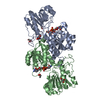
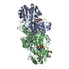
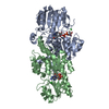
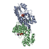
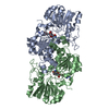
| Title | Crystal structure of formate dehydrogenase FDH2 D222A/Q223R enzyme from Granulicella mallensis MP5ACTX8 in complex with NADP and azide. | ||||||
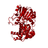
 Components Components | Formate dehydrogenase | ||||||
 Keywords Keywords | OXIDOREDUCTASE / formate dehydrogenase / NAD / NADP | ||||||
| Function / homology |  Function and homology information Function and homology informationformate catabolic process / formate dehydrogenase / formate dehydrogenase (NAD+) activity / oxidoreductase activity, acting on the CH-OH group of donors, NAD or NADP as acceptor / NAD binding / cytoplasm Similarity search - Function | ||||||
| Biological species |  Granulicella mallensis MP5ACTX8 (bacteria) Granulicella mallensis MP5ACTX8 (bacteria) | ||||||
| Method |  X-RAY DIFFRACTION / SYNCHROTRON / MOLECULAR REPLACEMENT / Resolution: 2.15 Å X-RAY DIFFRACTION / SYNCHROTRON / MOLECULAR REPLACEMENT / Resolution: 2.15 Å | ||||||
 Authors Authors | Robescu, M.S. / Rubini, R. / Filippini, F. / Bergantino, B. / Cendron, L. | ||||||
 Citation Citation | Journal: Chemcatchem / Year: 2020 Title: From the Amelioration of a NADP+-dependent Formate Dehydrogenase to the Discovery of a New Enzyme: Round Trip from Theory to Practice Authors: Robescu, M.S. / Rubini, R. / Beneventi, E. / Tavanti, M. / Lonigro, C. / Zito, F. / Filippini, F. / Cendron, L. / Bergantino, E. | ||||||
| History |
|
- Structure visualization
| Structure viewer | Molecule: MolmilJmol/JSmol |
|---|
- Downloads & links
-Download
| PDBx/mmCIF format | 6t9w.cif.gz | 322.9 KB | Display | PDBx/mmCIF format |
|---|---|---|---|---|
| PDB format | pdb6t9w.ent.gz | 261.3 KB | Display | PDB format |
| PDBx/mmJSON format | 6t9w.json.gz | Tree view | PDBx/mmJSON format | |
| Others |  Other downloads Other downloads |
-Validation report
| Arichive directory | https://data.pdbj.org/pub/pdb/validation_reports/t9/6t9wftp://data.pdbj.org/pub/pdb/validation_reports/t9/6t9w | HTTPS FTP |
|---|
-Related structure data
| Related structure data | 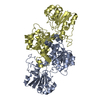 6t8cSC 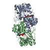 6t9xC 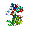 6tb6C S: Starting model for refinement C: citing same article ( |
|---|---|
| Similar structure data |


-Links
 PDBj
PDBj- Assembly
| Deposited unit | 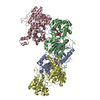
| ||||||||
|---|---|---|---|---|---|---|---|---|---|
| 1 | 
| ||||||||
| 2 | 
| ||||||||
| Unit cell |
|
-Components
| #1: Protein | Mass: 42574.621 Da / Num. of mol.: 4 Source method: isolated from a genetically manipulated source Details: not assigned coordinates correspond to not atoms not visible in the electron density Source: (gene. exp.) Granulicella mallensis MP5ACTX8 (bacteria)Gene: AciX8_0868 / Production host: #2: Chemical |   Mass: 743.405 Da / Num. of mol.: 3 / Source method: obtained synthetically / Formula: C21H28N7O17P3 / Feature type: SUBJECT OF INVESTIGATION Mass: 743.405 Da / Num. of mol.: 3 / Source method: obtained synthetically / Formula: C21H28N7O17P3 / Feature type: SUBJECT OF INVESTIGATION#3: Chemical |   Mass: 42.020 Da / Num. of mol.: 3 / Source method: obtained synthetically / Formula: N3 / Feature type: SUBJECT OF INVESTIGATION Mass: 42.020 Da / Num. of mol.: 3 / Source method: obtained synthetically / Formula: N3 / Feature type: SUBJECT OF INVESTIGATION#4: Water | ChemComp-HOH / |  Mass: 18.015 Da / Num. of mol.: 745 / Source method: isolated from a natural source / Formula: H2O Mass: 18.015 Da / Num. of mol.: 745 / Source method: isolated from a natural source / Formula: H2OHas ligand of interest | Y | |
|---|
-Experimental details
-Experiment
| Experiment | Method: X-RAY DIFFRACTION / Number of used crystals: 1 |
|---|
- Sample preparation
| Crystal | Density Matthews: 2.47 Å3/Da / Density % sol: 50.28 % |
|---|---|
| Crystal grow | Temperature: 293 K / Method: vapor diffusion, sitting drop / pH: 5 / Details: 0.1 M Citrate pH 5.0, 20 % w/v PEG 6000 |
-Data collection
| Diffraction | Mean temperature: 100 K / Serial crystal experiment: N |
|---|---|
| Diffraction source | Source: SYNCHROTRON / Site: ELETTRA  / Beamline: 11.2C / Wavelength: 0.98 Å / Beamline: 11.2C / Wavelength: 0.98 Å |
| Detector | Type: DECTRIS PILATUS3 S 6M / Detector: PIXEL / Date: Feb 7, 2019 |
| Radiation | Protocol: SINGLE WAVELENGTH / Monochromatic (M) / Laue (L): M / Scattering type: x-ray |
| Radiation wavelength | Wavelength: 0.98 Å / Relative weight: 1 |
| Reflection | Resolution: 2.15→70.65 Å / Num. obs: 153758 / % possible obs: 99.1 % / Redundancy: 4.6 % / CC1/2: 0.991 / Rmerge(I) obs: 0.109 / Net I/σ(I): 7.3 |
| Reflection shell | Resolution: 2.15→70.56 Å / Rmerge(I) obs: 0.709 / Mean I/σ(I) obs: 2 / Num. unique obs: 3923 / CC1/2: 0.683 |
- Processing
| Software |
| ||||||||||||||||||
|---|---|---|---|---|---|---|---|---|---|---|---|---|---|---|---|---|---|---|---|
| Refinement | Method to determine structure: MOLECULAR REPLACEMENT Starting model: 6T8C Resolution: 2.15→54.1 Å / Cross valid method: THROUGHOUT
| ||||||||||||||||||
| Displacement parameters | Biso max: 167.86 Å2 / Biso mean: 25.6723 Å2 / Biso min: 4.89 Å2 | ||||||||||||||||||
| Refinement step | Cycle: LAST / Resolution: 2.15→54.1 Å
|
