 Movie
Movie Controller


+ Open data

- Basic information
| Entry | Database: PDB / ID: 6t96 | ||||||
|---|---|---|---|---|---|---|---|
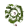
| Title | Photorhabdus laumondii subsp. laumondii lectin PLL3 | ||||||
 Components Components | Lectin PLL3 | ||||||
 Keywords Keywords | SUGAR BINDING PROTEIN / lectin / beta-propeller / carbohydrate-binding protein | ||||||
| Function / homology | : / PLL-like beta propeller / Photorhabdus luminescens subsp. laumondii TTO1 complete genome segment 3/17 Function and homology information Function and homology information | ||||||
| Biological species |  Photorhabdus laumondii subsp. laumondii TTO1 (bacteria) Photorhabdus laumondii subsp. laumondii TTO1 (bacteria) | ||||||
| Method |  X-RAY DIFFRACTION / SYNCHROTRON / MOLECULAR REPLACEMENT / Resolution: 1.65 Å X-RAY DIFFRACTION / SYNCHROTRON / MOLECULAR REPLACEMENT / Resolution: 1.65 Å | ||||||
 Authors Authors | Melicher, F. / Houser, J. / Fujdiarova, E. / Faltinek, L. / Wimmerova, M. | ||||||
 Citation Citation | Journal: Molecules / Year: 2019 Title: Lectin PLL3, a Novel Monomeric Member of the Seven-Bladed beta-Propeller Lectin Family. Authors: Faltinek, L. / Fujdiarova, E. / Melicher, F. / Houser, J. / Kasakova, M. / Kondakov, N. / Kononov, L. / Parkan, K. / Vidal, S. / Wimmerova, M. | ||||||
| History |
|
- Structure visualization
| Structure viewer | Molecule: MolmilJmol/JSmol |
|---|
- Downloads & links
-Download
| PDBx/mmCIF format | 6t96.cif.gz | 91.4 KB | Display | PDBx/mmCIF format |
|---|---|---|---|---|
| PDB format | pdb6t96.ent.gz | 65.9 KB | Display | PDB format |
| PDBx/mmJSON format | 6t96.json.gz | Tree view | PDBx/mmJSON format | |
| Others |  Other downloads Other downloads |
-Validation report
| Arichive directory | https://data.pdbj.org/pub/pdb/validation_reports/t9/6t96ftp://data.pdbj.org/pub/pdb/validation_reports/t9/6t96 | HTTPS FTP |
|---|
-Related structure data
| Related structure data | 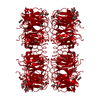 5c9pS S: Starting model for refinement |
|---|---|
| Similar structure data |


-Links
 PDBj
PDBj- Assembly
| Deposited unit | 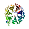
| ||||||||
|---|---|---|---|---|---|---|---|---|---|
| 1 |
| ||||||||
| Unit cell |
|
-Components
| #1: Protein | Mass: 40525.156 Da / Num. of mol.: 1 Source method: isolated from a genetically manipulated source Details: CYS at position 347 is found to be oxidized in the X-ray structure Source: (gene. exp.) Photorhabdus laumondii subsp. laumondii TTO1 (bacteria)Gene: plu0735 / Production host: |
|---|---|
| #2: Chemical | ChemComp-NA /   Mass: 22.990 Da / Num. of mol.: 1 / Source method: obtained synthetically / Formula: Na Mass: 22.990 Da / Num. of mol.: 1 / Source method: obtained synthetically / Formula: Na |
| #3: Water | ChemComp-HOH /  Mass: 18.015 Da / Num. of mol.: 325 / Source method: isolated from a natural source / Formula: H2O Mass: 18.015 Da / Num. of mol.: 325 / Source method: isolated from a natural source / Formula: H2O |
| Has ligand of interest | N |
| Has protein modification | Y |
-Experimental details
-Experiment
| Experiment | Method: X-RAY DIFFRACTION / Number of used crystals: 1 |
|---|
- Sample preparation
| Crystal | Density Matthews: 1.86 Å3/Da / Density % sol: 33.82 % |
|---|---|
| Crystal grow | Temperature: 293 K / Method: vapor diffusion, sitting drop / Details: 0.2 M NaF, 14% PEG 3350 |
-Data collection
| Diffraction | Mean temperature: 100 K / Serial crystal experiment: N |
|---|---|
| Diffraction source | Source: SYNCHROTRON / Site: BESSY  / Beamline: 14.1 / Wavelength: 0.9184 Å / Beamline: 14.1 / Wavelength: 0.9184 Å |
| Detector | Type: DECTRIS PILATUS3 6M / Detector: PIXEL / Date: Jun 15, 2019 |
| Radiation | Protocol: SINGLE WAVELENGTH / Monochromatic (M) / Laue (L): M / Scattering type: x-ray |
| Radiation wavelength | Wavelength: 0.9184 Å / Relative weight: 1 |
| Reflection | Resolution: 1.65→45.53 Å / Num. all: 37089 / Num. obs: 37089 / % possible obs: 99.9 % / Redundancy: 7.3 % / CC1/2: 0.997 / Rmerge(I) obs: 0.143 / Rpim(I) all: 0.057 / Rrim(I) all: 0.154 / Rsym value: 0.143 / Net I/σ(I): 8.2 / Num. measured all: 271406 |
| Reflection shell | Resolution: 1.65→1.74 Å / Redundancy: 7.4 % / Rmerge(I) obs: 0.989 / Num. unique obs: 5293 / CC1/2: 0.708 / Rpim(I) all: 0.024 / Rrim(I) all: 0.065 / Rsym value: 0.06 / % possible all: 99.2 |
- Processing
| Software |
| ||||||||||||||||||||||||||||||||||||||||||||||||||||||||||||
|---|---|---|---|---|---|---|---|---|---|---|---|---|---|---|---|---|---|---|---|---|---|---|---|---|---|---|---|---|---|---|---|---|---|---|---|---|---|---|---|---|---|---|---|---|---|---|---|---|---|---|---|---|---|---|---|---|---|---|---|---|---|
| Refinement | Method to determine structure: MOLECULAR REPLACEMENT Starting model: 5C9P Resolution: 1.65→45.53 Å / Cor.coef. Fo:Fc: 0.968 / Cor.coef. Fo:Fc free: 0.959 / SU B: 2.568 / SU ML: 0.082 / Cross valid method: THROUGHOUT / σ(F): 0 / ESU R: 0.105 / ESU R Free: 0.098 / Stereochemistry target values: MAXIMUM LIKELIHOOD Details: HYDROGENS HAVE BEEN ADDED IN THE RIDING POSITIONS U VALUES : REFINED INDIVIDUALLY
| ||||||||||||||||||||||||||||||||||||||||||||||||||||||||||||
| Solvent computation | Ion probe radii: 0.8 Å / Shrinkage radii: 0.8 Å / VDW probe radii: 1.2 Å / Solvent model: MASK | ||||||||||||||||||||||||||||||||||||||||||||||||||||||||||||
| Displacement parameters | Biso max: 70.58 Å2 / Biso mean: 18.29 Å2 / Biso min: 10.09 Å2
| ||||||||||||||||||||||||||||||||||||||||||||||||||||||||||||
| Refinement step | Cycle: final / Resolution: 1.65→45.53 Å
| ||||||||||||||||||||||||||||||||||||||||||||||||||||||||||||
| Refine LS restraints |
| ||||||||||||||||||||||||||||||||||||||||||||||||||||||||||||
| LS refinement shell | Resolution: 1.65→1.693 Å / Rfactor Rfree error: 0 / Total num. of bins used: 20
|
