 Movie
Movie Controller

[English] 日本語
 Yorodumi
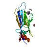
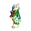
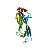
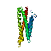
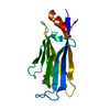
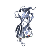
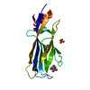
- PDB-6t1n: Crystal structure of MLLT1 (ENL) YEATS domain in complexed with b... -
+ Open data

- Basic information
| Entry | Database: PDB / ID: 6t1n | ||||||
|---|---|---|---|---|---|---|---|
| Title | Crystal structure of MLLT1 (ENL) YEATS domain in complexed with benzimidazole-amide derivative 5 | ||||||
 Components Components | Protein ENL | ||||||
 Keywords Keywords | TRANSCRIPTION / YEATS domain / ENL / MLLT1 / chemical probe / inhibitor / Structural Genomics / Structural Genomics Consortium / SGC | ||||||
| Function / homology |  Function and homology information Function and homology informationRNA Polymerase II Transcription Elongation / Formation of RNA Pol II elongation complex / RNA Polymerase II Pre-transcription Events / transcription elongation factor complex / fibrillar center / chromatin binding / positive regulation of DNA-templated transcription / nucleoplasm / cytosol Similarity search - Function | ||||||
| Biological species |  Homo sapiens (human) Homo sapiens (human) | ||||||
| Method |  X-RAY DIFFRACTION / SYNCHROTRON / MOLECULAR REPLACEMENT / Resolution: 1.95 Å X-RAY DIFFRACTION / SYNCHROTRON / MOLECULAR REPLACEMENT / Resolution: 1.95 Å | ||||||
 Authors Authors | Chaikuad, A. / Heidenreich, D. / Moustakim, M. / Arrowsmith, C.H. / Edwards, A.M. / Bountra, C. / Fedorov, O. / Brennan, P.E. / Knapp, S. / Structural Genomics Consortium (SGC) | ||||||
 Citation Citation | Journal: Acs Med.Chem.Lett. / Year: 2019 Title: Structural Insights into Interaction Mechanisms of Alternative Piperazine-urea YEATS Domain Binders in MLLT1. Authors: Ni, X. / Heidenreich, D. / Christott, T. / Bennett, J. / Moustakim, M. / Brennan, P.E. / Fedorov, O. / Knapp, S. / Chaikuad, A. | ||||||
| History |
|
- Structure visualization
| Structure viewer | Molecule: MolmilJmol/JSmol |
|---|
- Downloads & links
-Download
| PDBx/mmCIF format | 6t1n.cif.gz | 76.9 KB | Display | PDBx/mmCIF format |
|---|---|---|---|---|
| PDB format | pdb6t1n.ent.gz | 56.1 KB | Display | PDB format |
| PDBx/mmJSON format | 6t1n.json.gz | Tree view | PDBx/mmJSON format | |
| Others |  Other downloads Other downloads |
-Validation report
| Arichive directory | https://data.pdbj.org/pub/pdb/validation_reports/t1/6t1nftp://data.pdbj.org/pub/pdb/validation_reports/t1/6t1n | HTTPS FTP |
|---|
-Related structure data
| Related structure data | 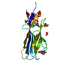 6t1iC 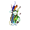 6t1jC 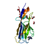 6t1lC  6t1mC 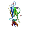 6t1oC 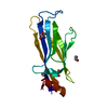 6hq0S S: Starting model for refinement C: citing same article ( |
|---|---|
| Similar structure data |


-Links
 PDBj
PDBj

- Assembly
| Deposited unit | 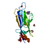
| ||||||||
|---|---|---|---|---|---|---|---|---|---|
| 1 |
| ||||||||
| Unit cell |
|
-Components
| #1: Protein | Mass: 18225.002 Da / Num. of mol.: 1 Source method: isolated from a genetically manipulated source Source: (gene. exp.) Homo sapiens (human) / Gene: MLLT1, ENL, LTG19, YEATS1 / Plasmid: pNIC-CH / Production host:  | ||||
|---|---|---|---|---|---|
| #2: Chemical | ChemComp-M7Z / 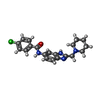  Mass: 368.860 Da / Num. of mol.: 1 / Source method: obtained synthetically / Formula: C20H21ClN4O / Feature type: SUBJECT OF INVESTIGATION Mass: 368.860 Da / Num. of mol.: 1 / Source method: obtained synthetically / Formula: C20H21ClN4O / Feature type: SUBJECT OF INVESTIGATION | ||||
| #3: Chemical | ChemComp-EDO /   Mass: 62.068 Da / Num. of mol.: 7 / Source method: obtained synthetically / Formula: C2H6O2 Mass: 62.068 Da / Num. of mol.: 7 / Source method: obtained synthetically / Formula: C2H6O2#4: Water | ChemComp-HOH / |  Mass: 18.015 Da / Num. of mol.: 71 / Source method: isolated from a natural source / Formula: H2O Mass: 18.015 Da / Num. of mol.: 71 / Source method: isolated from a natural source / Formula: H2OHas ligand of interest | Y | |
-Experimental details
-Experiment
| Experiment | Method: X-RAY DIFFRACTION / Number of used crystals: 1 |
|---|
- Sample preparation
| Crystal | Density Matthews: 2.18 Å3/Da / Density % sol: 43.63 % |
|---|---|
| Crystal grow | Temperature: 293.15 K / Method: vapor diffusion, sitting drop / pH: 5.5 Details: 25% PEG 3350, 0.2M sodium chloride, 0.1M bis-tris pH 5.5 |
-Data collection
| Diffraction | Mean temperature: 100 K / Serial crystal experiment: N | |||||||||||||||||||||||||||
|---|---|---|---|---|---|---|---|---|---|---|---|---|---|---|---|---|---|---|---|---|---|---|---|---|---|---|---|---|
| Diffraction source | Source: SYNCHROTRON / Site: SLS  / Beamline: X06SA / Wavelength: 1 Å / Beamline: X06SA / Wavelength: 1 Å | |||||||||||||||||||||||||||
| Detector | Type: DECTRIS EIGER X 16M / Detector: PIXEL / Date: Sep 21, 2018 | |||||||||||||||||||||||||||
| Radiation | Protocol: SINGLE WAVELENGTH / Monochromatic (M) / Laue (L): M / Scattering type: x-ray | |||||||||||||||||||||||||||
| Radiation wavelength | Wavelength: 1 Å / Relative weight: 1 | |||||||||||||||||||||||||||
| Reflection | Resolution: 1.95→49.01 Å / Num. obs: 12512 / % possible obs: 100 % / Redundancy: 7.1 % / Biso Wilson estimate: 33.6 Å2 / CC1/2: 0.997 / Rmerge(I) obs: 0.082 / Rpim(I) all: 0.033 / Rrim(I) all: 0.092 / Net I/σ(I): 9.8 | |||||||||||||||||||||||||||
| Reflection shell | Diffraction-ID: 1
|
- Processing
| Software |
| ||||||||||||||||||||||||||||||||||||||||||||||||||||||||||||
|---|---|---|---|---|---|---|---|---|---|---|---|---|---|---|---|---|---|---|---|---|---|---|---|---|---|---|---|---|---|---|---|---|---|---|---|---|---|---|---|---|---|---|---|---|---|---|---|---|---|---|---|---|---|---|---|---|---|---|---|---|---|
| Refinement | Method to determine structure: MOLECULAR REPLACEMENT Starting model: 6HQ0 Resolution: 1.95→45.97 Å / Cor.coef. Fo:Fc: 0.957 / Cor.coef. Fo:Fc free: 0.912 / SU B: 10.997 / SU ML: 0.152 / SU R Cruickshank DPI: 0.1825 / Cross valid method: THROUGHOUT / σ(F): 0 / ESU R: 0.182 / ESU R Free: 0.174 Details: U VALUES : WITH TLS ADDED HYDROGENS HAVE BEEN ADDED IN THE RIDING POSITIONS
| ||||||||||||||||||||||||||||||||||||||||||||||||||||||||||||
| Solvent computation | Ion probe radii: 0.8 Å / Shrinkage radii: 0.8 Å / VDW probe radii: 1.2 Å | ||||||||||||||||||||||||||||||||||||||||||||||||||||||||||||
| Displacement parameters | Biso max: 99.07 Å2 / Biso mean: 42.801 Å2 / Biso min: 25.57 Å2
| ||||||||||||||||||||||||||||||||||||||||||||||||||||||||||||
| Refinement step | Cycle: final / Resolution: 1.95→45.97 Å
| ||||||||||||||||||||||||||||||||||||||||||||||||||||||||||||
| Refine LS restraints |
| ||||||||||||||||||||||||||||||||||||||||||||||||||||||||||||
| LS refinement shell | Resolution: 1.95→2.001 Å / Rfactor Rfree error: 0 / Total num. of bins used: 20
| ||||||||||||||||||||||||||||||||||||||||||||||||||||||||||||
| Refinement TLS params. | Method: refined / Origin x: -0.6054 Å / Origin y: -3.8261 Å / Origin z: 12.7247 Å
|
