Mass: 18.015 Da / Num. of mol.: 160 / Source method: isolated from a natural source / Formula: H2O
Has ligand of interest
Y
Has protein modification
Y
-
Experimental details
-
Experiment
Experiment
Method: X-RAY DIFFRACTION / Number of used crystals: 1
-
Sample preparation
Crystal
Density Matthews: 2.08 Å3/Da / Density % sol: 40.96 % / Mosaicity: 0.26 °
Crystal grow
Temperature: 298 K / Method: vapor diffusion, hanging drop / pH: 7 Details: Crystals grew in 0.1 M sodium acetate, 0.3 M sodium chloride at pH 4.6 in the presence of 0.1 M Lithium sulfate. After growth, and before measurement, these crystals were soaked in the dye ...Details: Crystals grew in 0.1 M sodium acetate, 0.3 M sodium chloride at pH 4.6 in the presence of 0.1 M Lithium sulfate. After growth, and before measurement, these crystals were soaked in the dye solution: saturated bromophenol blue, 0.3 M sodium chloride, 100 mM Lithium sulfate, 100 mM phosphate buffer at pH 7.0
-
Data collection
Diffraction
Mean temperature: 100 K / Serial crystal experiment: N
Diffraction source
Source: SYNCHROTRON / Site: ALBA / Beamline: XALOC / Wavelength: 0.979 Å
Resolution: 0.97→19.6 Å / SU ML: 0.0969 / Cross valid method: FREE R-VALUE / σ(F): 1.91 / Phase error: 19.7886 Stereochemistry target values: GeoStd + Monomer Library + CDL v1.2 Details: The ligand geometry validation is not accurate because the molecule is a resonant form.
Rfactor
Num. reflection
% reflection
Rfree
0.1983
6932
5.06 %
Rwork
0.1731
130072
-
obs
0.1744
137004
98.87 %
Solvent computation
Shrinkage radii: 0.9 Å / VDW probe radii: 1.11 Å / Solvent model: FLAT BULK SOLVENT MODEL
Displacement parameters
Biso mean: 14.21 Å2
Refinement step
Cycle: LAST / Resolution: 0.97→19.6 Å
Protein
Nucleic acid
Ligand
Solvent
Total
Num. atoms
1001
0
59
160
1220
Refine LS restraints
Refine-ID
Type
Dev ideal
Number
X-RAY DIFFRACTION
f_bond_d
0.0093
1110
X-RAY DIFFRACTION
f_angle_d
1.0561
1514
X-RAY DIFFRACTION
f_chiral_restr
0.0768
150
X-RAY DIFFRACTION
f_plane_restr
0.0055
193
X-RAY DIFFRACTION
f_dihedral_angle_d
21.733
423
LS refinement shell
Resolution (Å)
Rfactor Rfree
Num. reflection Rfree
Rfactor Rwork
Num. reflection Rwork
Refine-ID
% reflection obs (%)
0.97-0.98
0.3655
233
0.3033
4177
X-RAY DIFFRACTION
95.25
0.98-0.99
0.2977
238
0.2768
4284
X-RAY DIFFRACTION
98.52
0.99-1
0.2854
265
0.2605
4297
X-RAY DIFFRACTION
97.94
1-1.01
0.2881
198
0.2429
4319
X-RAY DIFFRACTION
97.98
1.01-1.03
0.2369
214
0.2376
4356
X-RAY DIFFRACTION
99.15
1.03-1.04
0.2446
249
0.2265
4382
X-RAY DIFFRACTION
98.91
1.04-1.06
0.2442
220
0.21
4293
X-RAY DIFFRACTION
99.51
1.06-1.07
0.2155
206
0.1961
4402
X-RAY DIFFRACTION
99.25
1.07-1.09
0.2295
219
0.1879
4380
X-RAY DIFFRACTION
99.2
1.09-1.11
0.215
245
0.1804
4334
X-RAY DIFFRACTION
99.09
1.11-1.13
0.1747
215
0.1739
4379
X-RAY DIFFRACTION
99.24
1.13-1.15
0.1996
233
0.1661
4307
X-RAY DIFFRACTION
99.1
1.15-1.17
0.1876
255
0.1666
4371
X-RAY DIFFRACTION
99.1
1.17-1.19
0.1493
238
0.1593
4310
X-RAY DIFFRACTION
99.43
1.19-1.22
0.1697
226
0.1641
4355
X-RAY DIFFRACTION
99.31
1.22-1.25
0.1776
219
0.1706
4359
X-RAY DIFFRACTION
99.54
1.25-1.28
0.2158
240
0.1701
4364
X-RAY DIFFRACTION
99.2
1.28-1.31
0.1909
226
0.1697
4401
X-RAY DIFFRACTION
99.42
1.31-1.35
0.1927
265
0.1765
4269
X-RAY DIFFRACTION
99.32
1.35-1.39
0.1929
247
0.1752
4356
X-RAY DIFFRACTION
99.52
1.39-1.44
0.2088
220
0.1661
4402
X-RAY DIFFRACTION
99.57
1.44-1.5
0.1824
203
0.1656
4378
X-RAY DIFFRACTION
99.46
1.5-1.57
0.1694
196
0.1662
4396
X-RAY DIFFRACTION
99.5
1.57-1.65
0.1853
222
0.1626
4345
X-RAY DIFFRACTION
99.24
1.65-1.76
0.1891
261
0.1681
4316
X-RAY DIFFRACTION
98.43
1.76-1.89
0.1751
193
0.1703
4349
X-RAY DIFFRACTION
98.52
1.89-2.08
0.1878
239
0.1645
4280
X-RAY DIFFRACTION
98.35
2.08-2.38
0.1975
257
0.1675
4306
X-RAY DIFFRACTION
98.49
2.38-3
0.1833
209
0.1673
4346
X-RAY DIFFRACTION
98.32
3-19.6
0.2134
281
0.1726
4259
X-RAY DIFFRACTION
98.57
+
About Yorodumi
-
News
-
Feb 9, 2022. New format data for meta-information of EMDB entries
New format data for meta-information of EMDB entries
Version 3 of the EMDB header file is now the official format.
The previous official version 1.9 will be removed from the archive.
In the structure databanks used in Yorodumi, some data are registered as the other names, "COVID-19 virus" and "2019-nCoV". Here are the details of the virus and the list of structure data.
Jan 31, 2019. EMDB accession codes are about to change! (news from PDBe EMDB page)
EMDB accession codes are about to change! (news from PDBe EMDB page)
The allocation of 4 digits for EMDB accession codes will soon come to an end. Whilst these codes will remain in use, new EMDB accession codes will include an additional digit and will expand incrementally as the available range of codes is exhausted. The current 4-digit format prefixed with “EMD-” (i.e. EMD-XXXX) will advance to a 5-digit format (i.e. EMD-XXXXX), and so on. It is currently estimated that the 4-digit codes will be depleted around Spring 2019, at which point the 5-digit format will come into force.
The EM Navigator/Yorodumi systems omit the EMD- prefix.
Related info.:Q: What is EMD? / ID/Accession-code notation in Yorodumi/EM Navigator
Yorodumi is a browser for structure data from EMDB, PDB, SASBDB, etc.
This page is also the successor to EM Navigator detail page, and also detail information page/front-end page for Omokage search.
The word "yorodu" (or yorozu) is an old Japanese word meaning "ten thousand". "mi" (miru) is to see.
Related info.:EMDB / PDB / SASBDB / Comparison of 3 databanks / Yorodumi Search / Aug 31, 2016. New EM Navigator & Yorodumi / Yorodumi Papers / Jmol/JSmol / Function and homology information / Changes in new EM Navigator and Yorodumi
 Movie
Movie Controller

 Yorodumi
 Open data
 Basic information
 Components
 Keywords
 Function and homology information
 X-RAY DIFFRACTION /
  Authors
 Spain, 1items
  Citation
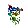
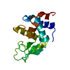
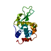
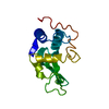
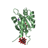
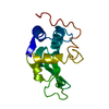
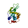
 Structure visualization
 Downloads & links
 Other downloads
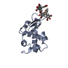


 PDBj
PDBj


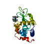
 Assembly
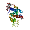

 Mass: 35.453 Da / Num. of mol.: 1 / Source method: obtained synthetically / Formula: Cl
 Mass: 669.961 Da / Num. of mol.: 2 / Source method: obtained synthetically / Formula: C19H10Br4O5S / Feature type: SUBJECT OF INVESTIGATION
 Mass: 18.015 Da / Num. of mol.: 160 / Source method: isolated from a natural source / Formula: H2O
 Sample preparation
 Processing