 Movie
Movie Controller

[English] 日本語
 Yorodumi
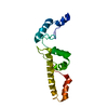
- PDB-6stc: Crystal structure of the tick chemokine-binding protein Evasin-4 ... -
+ Open data

- Basic information
| Entry | Database: PDB / ID: 6stc | ||||||
|---|---|---|---|---|---|---|---|
| Title | Crystal structure of the tick chemokine-binding protein Evasin-4 (SG 2) | ||||||
 Components Components | Evasin-4 | ||||||
 Keywords Keywords | IMMUNE SYSTEM / CHEMOKINE-BINDING PROTEIN / TICKS | ||||||
| Function / homology | negative regulation of chemokine activity / Evasins Class A / Evasins Class A / C-C chemokine binding / Immunoglobulin-like domain / extracellular region / ACETATE ION / DI(HYDROXYETHYL)ETHER / Evasin-4 Function and homology information Function and homology information | ||||||
| Biological species |  Rhipicephalus sanguineus (brown dog tick) Rhipicephalus sanguineus (brown dog tick) | ||||||
| Method |  X-RAY DIFFRACTION / SYNCHROTRON / MOLECULAR REPLACEMENT / Resolution: 1.69 Å X-RAY DIFFRACTION / SYNCHROTRON / MOLECULAR REPLACEMENT / Resolution: 1.69 Å | ||||||
 Authors Authors | Ramirez-Escudero, M. / Janssen, B.J.C. | ||||||
| Funding support | 1items
| ||||||
 Citation Citation | Journal: J.Biol.Chem. / Year: 2020 Title: Structural characterization of anti-CCL5 activity of the tick salivary protein evasin-4. Authors: Denisov, S.S. / Ramirez-Escudero, M. / Heinzmann, A.C.A. / Ippel, J.H. / Dawson, P.E. / Koenen, R.R. / Hackeng, T.M. / Janssen, B.J.C. / Dijkgraaf, I. | ||||||
| History |
|
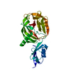
- Structure visualization
| Structure viewer | Molecule: MolmilJmol/JSmol |
|---|
- Downloads & links
-Download
| PDBx/mmCIF format | 6stc.cif.gz | 51.9 KB | Display | PDBx/mmCIF format |
|---|---|---|---|---|
| PDB format | pdb6stc.ent.gz | 35.9 KB | Display | PDB format |
| PDBx/mmJSON format | 6stc.json.gz | Tree view | PDBx/mmJSON format | |
| Others |  Other downloads Other downloads |
-Validation report
| Arichive directory | https://data.pdbj.org/pub/pdb/validation_reports/st/6stcftp://data.pdbj.org/pub/pdb/validation_reports/st/6stc | HTTPS FTP |
|---|
-Related structure data
| Related structure data | 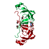 6st4SC 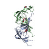 6steC 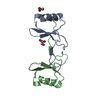 6stkC S: Starting model for refinement C: citing same article ( |
|---|---|
| Similar structure data |

-Links
 PDBj
PDBj- Assembly
| Deposited unit | 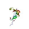
| ||||||||
|---|---|---|---|---|---|---|---|---|---|
| 1 | 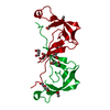
| ||||||||
| Unit cell |
|
-Components
| #1: Protein | Mass: 11316.322 Da / Num. of mol.: 1 Source method: isolated from a genetically manipulated source Source: (gene. exp.) Rhipicephalus sanguineus (brown dog tick)Production host:  | ||||||||
|---|---|---|---|---|---|---|---|---|---|
| #2: Chemical |   Mass: 59.044 Da / Num. of mol.: 3 / Source method: obtained synthetically / Formula: C2H3O2 Mass: 59.044 Da / Num. of mol.: 3 / Source method: obtained synthetically / Formula: C2H3O2#3: Chemical |   Mass: 106.120 Da / Num. of mol.: 3 / Source method: obtained synthetically / Formula: C4H10O3 Mass: 106.120 Da / Num. of mol.: 3 / Source method: obtained synthetically / Formula: C4H10O3#4: Water | ChemComp-HOH / |  Mass: 18.015 Da / Num. of mol.: 49 / Source method: isolated from a natural source / Formula: H2O Mass: 18.015 Da / Num. of mol.: 49 / Source method: isolated from a natural source / Formula: H2OHas ligand of interest | N | Has protein modification | Y | |
-Experimental details
-Experiment
| Experiment | Method: X-RAY DIFFRACTION / Number of used crystals: 1 |
|---|
- Sample preparation
| Crystal | Density Matthews: 2.77 Å3/Da / Density % sol: 55.66 % / Description: hexagonal prism |
|---|---|
| Crystal grow | Temperature: 291 K / Method: vapor diffusion, sitting drop / Details: 40% PEG 400, 0.1 M NaAc pH 4.5 |
-Data collection
| Diffraction | Mean temperature: 100 K / Serial crystal experiment: N |
|---|---|
| Diffraction source | Source: SYNCHROTRON / Site: ESRF  / Beamline: MASSIF-3 / Wavelength: 0.9677 Å / Beamline: MASSIF-3 / Wavelength: 0.9677 Å |
| Detector | Type: DECTRIS EIGER X 4M / Detector: PIXEL / Date: Aug 29, 2018 |
| Radiation | Protocol: SINGLE WAVELENGTH / Monochromatic (M) / Laue (L): M / Scattering type: x-ray |
| Radiation wavelength | Wavelength: 0.9677 Å / Relative weight: 1 |
| Reflection | Resolution: 1.69→43.06 Å / Num. obs: 14083 / % possible obs: 98.3 % / Redundancy: 3.8 % / CC1/2: 0.999 / Rmerge(I) obs: 0.054 / Net I/σ(I): 11.7 |
| Reflection shell | Resolution: 1.69→1.72 Å / Rmerge(I) obs: 0.928 / Mean I/σ(I) obs: 1.4 / Num. unique obs: 722 / CC1/2: 0.496 |
- Processing
| Software |
| ||||||||||||||||||||||||||||||||||||||||||||||||||||||||||||
|---|---|---|---|---|---|---|---|---|---|---|---|---|---|---|---|---|---|---|---|---|---|---|---|---|---|---|---|---|---|---|---|---|---|---|---|---|---|---|---|---|---|---|---|---|---|---|---|---|---|---|---|---|---|---|---|---|---|---|---|---|---|
| Refinement | Method to determine structure: MOLECULAR REPLACEMENT Starting model: 6ST4 Resolution: 1.69→30.79 Å / Cor.coef. Fo:Fc: 0.971 / Cor.coef. Fo:Fc free: 0.964 / SU B: 3.99 / SU ML: 0.06 / Cross valid method: THROUGHOUT / σ(F): 0 / ESU R: 0.083 / ESU R Free: 0.082 Details: HYDROGENS HAVE BEEN ADDED IN THE RIDING POSITIONS U VALUES : WITH TLS ADDED
| ||||||||||||||||||||||||||||||||||||||||||||||||||||||||||||
| Solvent computation | Ion probe radii: 0.8 Å / Shrinkage radii: 0.8 Å / VDW probe radii: 1.2 Å | ||||||||||||||||||||||||||||||||||||||||||||||||||||||||||||
| Displacement parameters | Biso max: 83.19 Å2 / Biso mean: 32.411 Å2 / Biso min: 22.03 Å2
| ||||||||||||||||||||||||||||||||||||||||||||||||||||||||||||
| Refinement step | Cycle: final / Resolution: 1.69→30.79 Å
| ||||||||||||||||||||||||||||||||||||||||||||||||||||||||||||
| Refine LS restraints |
| ||||||||||||||||||||||||||||||||||||||||||||||||||||||||||||
| LS refinement shell | Resolution: 1.69→1.734 Å / Rfactor Rfree error: 0 / Total num. of bins used: 20
| ||||||||||||||||||||||||||||||||||||||||||||||||||||||||||||
| Refinement TLS params. | Method: refined / Origin x: -26.639 Å / Origin y: 25.688 Å / Origin z: 9.112 Å
|
