 Movie
Movie Controller

[English] 日本語
 Yorodumi
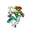
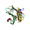
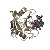
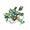
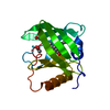
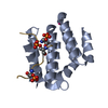
- PDB-6srt: Endolysine N-acetylmuramoyl-L-alanine amidase LysCS from Clostrid... -
+ Open data

- Basic information
| Entry | Database: PDB / ID: 6srt | ||||||
|---|---|---|---|---|---|---|---|
| Title | Endolysine N-acetylmuramoyl-L-alanine amidase LysCS from Clostridium intestinale URNW | ||||||
 Components Components | N-acetylmuramoyl-L-alanine amidase | ||||||
 Keywords Keywords | HYDROLASE / endolysine / zinc binding / amidase / cell wall degradation | ||||||
| Function / homology |  Function and homology information Function and homology informationN-acetylmuramoyl-L-alanine amidase activity / peptidoglycan catabolic process / zinc ion binding Similarity search - Function | ||||||
| Biological species |  Clostridium intestinale URNW (bacteria) Clostridium intestinale URNW (bacteria) | ||||||
| Method |  X-RAY DIFFRACTION / SYNCHROTRON / MOLECULAR REPLACEMENT / Resolution: 1.21 Å X-RAY DIFFRACTION / SYNCHROTRON / MOLECULAR REPLACEMENT / Resolution: 1.21 Å | ||||||
 Authors Authors | Hakansson, M. / Al-Karadaghi, S. / Plotka, M. / Kaczorowska, A.-K. / Kaczorowski, T. | ||||||
| Funding support |  Sweden, 1items Sweden, 1items
| ||||||
 Citation Citation | Journal: To Be Published Title: Structure and function of endolysines LysCS, LysC from Clostridium intestinale Authors: Plotka, M. / Hakansson, M. | ||||||
| History |
|
- Structure visualization
| Structure viewer | Molecule: MolmilJmol/JSmol |
|---|
- Downloads & links
-Download
| PDBx/mmCIF format | 6srt.cif.gz | 91.3 KB | Display | PDBx/mmCIF format |
|---|---|---|---|---|
| PDB format | pdb6srt.ent.gz | 68.3 KB | Display | PDB format |
| PDBx/mmJSON format | 6srt.json.gz | Tree view | PDBx/mmJSON format | |
| Others |  Other downloads Other downloads |
-Validation report
| Arichive directory | https://data.pdbj.org/pub/pdb/validation_reports/sr/6srtftp://data.pdbj.org/pub/pdb/validation_reports/sr/6srt | HTTPS FTP |
|---|
-Related structure data
| Related structure data |  6su5S S: Starting model for refinement |
|---|---|
| Similar structure data |


-Links
 PDBj
PDBj- Assembly
| Deposited unit | 
| ||||||||
|---|---|---|---|---|---|---|---|---|---|
| 1 |
| ||||||||
| Unit cell |
|
-Components
| #1: Protein | Mass: 19401.857 Da / Num. of mol.: 1 Source method: isolated from a genetically manipulated source Source: (gene. exp.) Clostridium intestinale URNW (bacteria)Gene: CINTURNW_1763 / Production host: | ||||||
|---|---|---|---|---|---|---|---|
| #2: Chemical | ChemComp-ZN /   Mass: 65.409 Da / Num. of mol.: 1 / Source method: obtained synthetically / Formula: Zn / Feature type: SUBJECT OF INVESTIGATION Mass: 65.409 Da / Num. of mol.: 1 / Source method: obtained synthetically / Formula: Zn / Feature type: SUBJECT OF INVESTIGATION | ||||||
| #3: Chemical |   Mass: 92.094 Da / Num. of mol.: 2 / Source method: obtained synthetically / Formula: C3H8O3 Mass: 92.094 Da / Num. of mol.: 2 / Source method: obtained synthetically / Formula: C3H8O3#4: Chemical |   Mass: 94.971 Da / Num. of mol.: 2 / Source method: obtained synthetically / Formula: PO4 / Feature type: SUBJECT OF INVESTIGATION Mass: 94.971 Da / Num. of mol.: 2 / Source method: obtained synthetically / Formula: PO4 / Feature type: SUBJECT OF INVESTIGATION#5: Water | ChemComp-HOH / |  Mass: 18.015 Da / Num. of mol.: 217 / Source method: isolated from a natural source / Formula: H2O Mass: 18.015 Da / Num. of mol.: 217 / Source method: isolated from a natural source / Formula: H2OHas ligand of interest | Y | |
-Experimental details
-Experiment
| Experiment | Method: X-RAY DIFFRACTION / Number of used crystals: 1 |
|---|
- Sample preparation
| Crystal | Density Matthews: 2.22 Å3/Da / Density % sol: 44.48 % |
|---|---|
| Crystal grow | Temperature: 293 K / Method: evaporation / pH: 7.4 / Details: NaKHPO4, PEG 3350 |
-Data collection
| Diffraction | Mean temperature: 100 K / Serial crystal experiment: N |
|---|---|
| Diffraction source | Source: SYNCHROTRON / Site: Diamond  / Beamline: I04 / Wavelength: 0.97951 Å / Beamline: I04 / Wavelength: 0.97951 Å |
| Detector | Type: DECTRIS PILATUS3 6M / Detector: PIXEL / Date: Apr 19, 2018 |
| Radiation | Protocol: SINGLE WAVELENGTH / Monochromatic (M) / Laue (L): M / Scattering type: x-ray |
| Radiation wavelength | Wavelength: 0.97951 Å / Relative weight: 1 |
| Reflection | Resolution: 1.21→44.2 Å / Num. obs: 48199 / % possible obs: 99.9 % / Redundancy: 5.2 % / CC1/2: 1 / Rmerge(I) obs: 0.078 / Net I/σ(I): 9.3 |
| Reflection shell | Resolution: 1.21→1.23 Å / Rmerge(I) obs: 1.22 / Mean I/σ(I) obs: 1.1 / Num. unique obs: 2370 / CC1/2: 0.5 |
- Processing
| Software |
| |||||||||||||||||||||||||||||||||||||||||||||||||||||||||||||||||
|---|---|---|---|---|---|---|---|---|---|---|---|---|---|---|---|---|---|---|---|---|---|---|---|---|---|---|---|---|---|---|---|---|---|---|---|---|---|---|---|---|---|---|---|---|---|---|---|---|---|---|---|---|---|---|---|---|---|---|---|---|---|---|---|---|---|---|
| Refinement | Method to determine structure: MOLECULAR REPLACEMENT Starting model: 6SU5 Resolution: 1.21→44.2 Å / Cor.coef. Fo:Fc: 0.985 / Cor.coef. Fo:Fc free: 0.977 / SU B: 1.184 / SU ML: 0.023 / Cross valid method: THROUGHOUT / σ(F): 0 / ESU R: 0.032 / ESU R Free: 0.036 Details: HYDROGENS HAVE BEEN ADDED IN THE RIDING POSITIONS U VALUES : REFINED INDIVIDUALLY
| |||||||||||||||||||||||||||||||||||||||||||||||||||||||||||||||||
| Solvent computation | Ion probe radii: 0.8 Å / Shrinkage radii: 0.8 Å / VDW probe radii: 1.2 Å | |||||||||||||||||||||||||||||||||||||||||||||||||||||||||||||||||
| Displacement parameters | Biso max: 93.18 Å2 / Biso mean: 16.913 Å2 / Biso min: 8.55 Å2
| |||||||||||||||||||||||||||||||||||||||||||||||||||||||||||||||||
| Refinement step | Cycle: final / Resolution: 1.21→44.2 Å
| |||||||||||||||||||||||||||||||||||||||||||||||||||||||||||||||||
| Refine LS restraints |
| |||||||||||||||||||||||||||||||||||||||||||||||||||||||||||||||||
| LS refinement shell | Resolution: 1.21→1.241 Å / Rfactor Rfree error: 0 / Total num. of bins used: 20
|
