 Movie
Movie Controller

[English] 日本語
 Yorodumi
- PDB-6sr6: Crystal structure of the RAC core with a pseudo substrate bound t... -
+ Open data

- Basic information
| Entry | Database: PDB / ID: 6sr6 | |||||||||
|---|---|---|---|---|---|---|---|---|---|---|
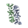
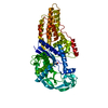
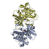
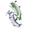
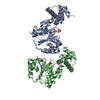
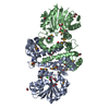
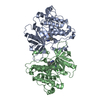
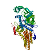
| Title | Crystal structure of the RAC core with a pseudo substrate bound to Ssz1 SBD | |||||||||
 Components Components |
| |||||||||
 Keywords Keywords | CHAPERONE / Hsp70 | |||||||||
| Function / homology |  Function and homology information Function and homology information'de novo' cotranslational protein folding / regulation of translational fidelity / Hsp70 protein binding / ATP-dependent protein folding chaperone / ribosome binding / ATP binding / nucleus / cytosol Similarity search - Function | |||||||||
| Biological species |  Chaetomium thermophilum (fungus) Chaetomium thermophilum (fungus) | |||||||||
| Method |  X-RAY DIFFRACTION / SYNCHROTRON / MOLECULAR REPLACEMENT / Resolution: 2.5 Å X-RAY DIFFRACTION / SYNCHROTRON / MOLECULAR REPLACEMENT / Resolution: 2.5 Å | |||||||||
 Authors Authors | Valentin Gese, G. / Lapouge, K. / Kopp, J. / Sinning, I. | |||||||||
| Funding support |  Germany, 2items Germany, 2items
| |||||||||
 Citation Citation | Journal: Nat Commun / Year: 2020 Title: The ribosome-associated complex RAC serves in a relay that directs nascent chains to Ssb. Authors: Zhang, Y. / Valentin Gese, G. / Conz, C. / Lapouge, K. / Kopp, J. / Wolfle, T. / Rospert, S. / Sinning, I. | |||||||||
| History |
|
- Structure visualization
| Structure viewer | Molecule: MolmilJmol/JSmol |
|---|
- Downloads & links
-Download
| PDBx/mmCIF format | 6sr6.cif.gz | 453.6 KB | Display | PDBx/mmCIF format |
|---|---|---|---|---|
| PDB format | pdb6sr6.ent.gz | 368.7 KB | Display | PDB format |
| PDBx/mmJSON format | 6sr6.json.gz | Tree view | PDBx/mmJSON format | |
| Others |  Other downloads Other downloads |
-Validation report
| Arichive directory | https://data.pdbj.org/pub/pdb/validation_reports/sr/6sr6ftp://data.pdbj.org/pub/pdb/validation_reports/sr/6sr6 | HTTPS FTP |
|---|
-Related structure data
| Related structure data |  5mb9S S: Starting model for refinement |
|---|---|
| Similar structure data |


-Links
 PDBj
PDBj


- Assembly
| Deposited unit | 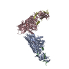
| ||||||||||
|---|---|---|---|---|---|---|---|---|---|---|---|
| 1 | 
| ||||||||||
| 2 | 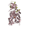
| ||||||||||
| Unit cell |
|
-Components
| #1: Protein | Mass: 63706.820 Da / Num. of mol.: 2 Source method: isolated from a genetically manipulated source Source: (gene. exp.) Chaetomium thermophilum (strain DSM 1495 / CBS 144.50 / IMI 039719) (fungus)Strain: DSM 1495 / CBS 144.50 / IMI 039719 / Gene: CTHT_0008010 / Production host:  #2: Protein | Mass: 6906.990 Da / Num. of mol.: 2 Source method: isolated from a genetically manipulated source Source: (gene. exp.) Chaetomium thermophilum (strain DSM 1495 / CBS 144.50 / IMI 039719) (fungus)Strain: DSM 1495 / CBS 144.50 / IMI 039719 / Gene: CTHT_0006310 / Production host: #3: Chemical |   Mass: 507.181 Da / Num. of mol.: 2 / Source method: obtained synthetically / Formula: C10H16N5O13P3 / Comment: ATP, energy-carrying molecule*YM Mass: 507.181 Da / Num. of mol.: 2 / Source method: obtained synthetically / Formula: C10H16N5O13P3 / Comment: ATP, energy-carrying molecule*YM#4: Chemical |   Mass: 24.305 Da / Num. of mol.: 2 / Source method: obtained synthetically / Formula: Mg Mass: 24.305 Da / Num. of mol.: 2 / Source method: obtained synthetically / Formula: Mg#5: Water | ChemComp-HOH / |  Mass: 18.015 Da / Num. of mol.: 91 / Source method: isolated from a natural source / Formula: H2O Mass: 18.015 Da / Num. of mol.: 91 / Source method: isolated from a natural source / Formula: H2OHas ligand of interest | N | |
|---|
-Experimental details
-Experiment
| Experiment | Method: X-RAY DIFFRACTION / Number of used crystals: 1 |
|---|
- Sample preparation
| Crystal | Density Matthews: 2.83 Å3/Da / Density % sol: 56.48 % |
|---|---|
| Crystal grow | Temperature: 291 K / Method: vapor diffusion, sitting drop / pH: 7.5 / Details: 20.5% (v/v) PEG 3350, 0.2 M ammonium acetate |
-Data collection
| Diffraction | Mean temperature: 100 K / Serial crystal experiment: N |
|---|---|
| Diffraction source | Source: SYNCHROTRON / Site: ESRF  / Beamline: MASSIF-3 / Wavelength: 0.9677 Å / Beamline: MASSIF-3 / Wavelength: 0.9677 Å |
| Detector | Type: DECTRIS EIGER X 4M / Detector: PIXEL / Date: May 5, 2018 |
| Radiation | Protocol: SINGLE WAVELENGTH / Monochromatic (M) / Laue (L): M / Scattering type: x-ray |
| Radiation wavelength | Wavelength: 0.9677 Å / Relative weight: 1 |
| Reflection | Resolution: 2.5→48.4 Å / Num. obs: 46860 / % possible obs: 98.9 % / Redundancy: 6 % / Biso Wilson estimate: 38.97 Å2 / Rmerge(I) obs: 0.134 / Net I/σ(I): 9.6 |
| Reflection shell | Resolution: 2.5→2.59 Å / Rmerge(I) obs: 0.706 / Mean I/σ(I) obs: 2.2 / Num. unique obs: 4516 / CC1/2: 0.658 |
- Processing
| Software |
| ||||||||||||||||||||
|---|---|---|---|---|---|---|---|---|---|---|---|---|---|---|---|---|---|---|---|---|---|
| Refinement | Method to determine structure: MOLECULAR REPLACEMENT Starting model: 5MB9 Resolution: 2.5→33.71 Å / Cross valid method: FREE R-VALUE
| ||||||||||||||||||||
| Refinement step | Cycle: LAST / Resolution: 2.5→33.71 Å
|
